Alumni Profile: Desmond Edwards, SB ’22

An interest in translating medicine for a wider audience

School of Science
February 6, 2025

Growing up hearing both English and Patois in rural Jamaica, he always had an interest in understanding other languages, so he studied French in high school and minored in it at MIT. As a child with persistent illnesses, he was frustrated that doctors couldn’t explain the “how” and “why” of what was happening in his body. “I wanted to understand how an entity so small that we can’t even see it with most microscopes is able to get into a massively intricate human body and completely shut it down in a matter of days,” he says.

Edwards, now an MIT graduate and a PhD candidate in microbiology and immunology at Stanford University—with a deferred MD admission in hand as well—feels closer to understanding things. The financial support he received at MIT from the Class of 1975 Scholarship Fund, he says, was one major reason that he chose MIT.

Support for research and discovery

I took a three-week Independent Activities Period boot camp designed to expose first-years with little or no research background to basic molecular biology and microbiology techniques. We had guidance from the professor and teaching assistants, but it was up to us what path we took. That intellectual freedom was part of what made me fall in love with academic research. The lecturer, Mandana Sassanfar, made it her personal mission to connect interested students to Undergraduate Research Opportunities Program placements, which is how I found myself in Professor Rebecca Lamason’s lab.

At the end of my first year, I debated whether to prioritize my academic research projects or leave for a higher-paying summer internship. My lab helped me apply for the Peter J. Eloranta Summer Undergraduate Research Fellowship, which provided funding that allowed me to stay for the summer, and I ended up staying in the lab for the rest of my time at MIT. One paper I coauthored (about developing new genetic tools to control pathogenic bacteria’s gene expression) was published this year.

French connections

French is one of the working languages of many global health programs, and being able to read documents in their original language has been helpful because many diseases that I care about impact Francophone countries like those in sub-Saharan and west Africa. In one French class, we had to analyze an original primary historical text, so I was able to look at an outbreak of plague in the 18th century and compare their public health response with ours to Covid-19. My MIT French classes have been useful in some very cool ways that I did not anticipate.

Translating medicine for the masses

When I go home and talk about my research, I often adapt folk stories, analogies, and relatable everyday situations to get points across since there might not be exact Patois words or phrases to directly convey what I’m describing. Taking these scientific concepts and breaking them all into bite-size pieces is important for the general American public too. I want to lead a scientific career that not only advances our understanding and treatment of infectious diseases, but also positively impacts policy, education, and outreach. Right now, this looks like a combination of being an academic/medical professor and eventually leading the Centers for Disease Control and Prevention.

Alumni Profile: Matthew Dolan, SB ’81

From Bench to Bedside and Beyond

Lillian Eden | Department of Biology
January 16, 2025

Matthew Dolan, SB ‘81, worked in the U.S. and abroad during a fascinating time in the field of immunology and virology.

In medical school, Matthew Dolan, SB ‘81, briefly considered specializing in orthopedic surgery because of the materials science nature of the work — but he soon realized that he didn’t have the innate skills required for that type of work. 

“I’ll be honest with you — I can’t parallel park,” he jokes. “You can consider a lot of things, but if you find the things that you’re good at and that excite you, you can hopefully move forward with those.” 

Dolan certainly has, tackling problems from bench to bedside and beyond. Both in the U.S. and abroad through the Air Force, Dolan has emerged as a leader in immunology and virology, and has served as Director of the Defense Institute for Medical Operations. He’s worked on everything from foodborne illnesses and Ebola to biological weapons and COVID-19, and has even been a guest speaker on NPR’s Science Friday

“This is fun and interesting, and I believe that, and I work hard to convey that — and it’s contagious,” he says. “You can affect people with that excitement.” 

Pieces of the Puzzle

Dolan fondly recalls his years at MIT, and is still in touch with many of the “brilliant” and “interesting” friends he made while in Cambridge. 

He notes that the challenges that were the most rewarding in his career were also the ones that MIT had uniquely prepared him for. Dolan, a Course 7 major, naturally took many classes outside of Biology as part of his undergraduate studies: organic chemistry was foundational for understanding toxicology while studying chemical weapons, while pathogens like Legionella, which causes pneumonia and can spread through water systems like ice machines or air conditioners, are solved at the interface between public health and ecology.

Man sitting on couch next to white dog with pointy ears.
Matthew Dolan stateside with his German Shepherd Sophie. Photo courtesy of Matthew Dolan.

“I learned that learning can be a high-intensity experience,” Dolan recalls. “You can be aggressive in your learning; you can learn and excel in a wide variety of things and gather up all the knowledge and knowledgeable people to work together towards solutions.”

Dolan, for example, worked in the Amazon Basin in Peru on a public health crisis of a sharp rise in childhood mortality due to malaria. The cause was a few degrees removed from the immediate problem: human agriculture had affected the Amazon’s tributaries, leading to still and stagnant water where before there had been rushing streams and rivers. This change in the environment allowed a certain mosquito species of “avid human biters” to thrive.  

“It can be helpful and important for some people to have a really comprehensive and contextual view of scientific problems and biological problems,” he says. “It’s very rewarding to put the pieces in a puzzle like that together.” 

Choosing To Serve

Dolan says a key to finding meaning in his work, especially during difficult times, is a sentiment from Alsatian polymath and Nobel Peace Prize winner Albert Schweitzer: “The only ones among you who will be really happy are those who will have sought and found how to serve.”

One of Dolan’s early formative experiences was working in the heart of the HIV/AIDS epidemic, at a time when there was no effective treatment. No matter how hard he worked, the patients would still die. 

“Failure is not an option — unless you have to fail. You can’t let the failures destroy you,” he says. “There are a lot of other battles out there, and it’s self-indulgent to ignore them and focus on your woe.” 

Lasting Impacts

Dolan couldn’t pick a favorite country, but notes that he’s always impressed seeing how people value the chance to excel with science and medicine when offered resources and respect. Ultimately, everyone he’s worked with, no matter their differences, was committed to solving problems and improving lives. 

Dolan worked in Russia after the Berlin Wall fell, on HIV/AIDS in Moscow and Tuberculosis in the Russian Far East. Although relations with Russia are currently tense, to say the least, Dolan remains optimistic for a brighter future. 

“People that were staunch adversaries can go on to do well together,” he says. “Sometimes, peace leads to partnership. Remembering that it was once possible gives me great hope.” 

Dolan understands that the most lasting impact he has had is, likely, teaching: time marches on, and discoveries can be lost to history, but teaching and training people continues and propagates. In addition to guiding the next generation of healthcare specialists, Dolan also developed programs in laboratory biosafety and biosecurity with the State Department and the Defense Department, and taught those programs around the world. 

“Working in prevention gives you the chance to take care of process problems before they become people problems — patient care problems,” he says. “I have been so impressed with the courageous and giving people that have worked with me.” 

From Molecules to Memory

On a biological foundation of ions and proteins, the brain forms, stores, and retrieves memories to inform intelligent behavior.

Noah Daly | Department of Biology
December 23, 2024

Whenever you go out to a restaurant to celebrate, your brain retrieves memories while forming new ones. You notice the room is elegant, that you’re surrounded by people you love, having meaningful conversations, and doing it all with good manners. Encoding these precious moments (and not barking at your waiter, expecting dessert before your appetizer), you rely heavily on plasticity, the ability of neurons to change the strength and quantity of their connections in response to new information or activity. The very existence of memory and our ability to retrieve it to guide our intelligent behavior are hypothesized to be movements of a neuroplastic symphony, manifested through chemical processes occurring across vast, interconnected networks of neurons.

During infancy, brain connectivity grows exponentially, rapidly increasing the number of synapses between neurons, some of which are then pruned back to select the most salient for optimal performance. This exuberant growth followed by experience-dependent optimization lays a foundation of connections to produce a functional brain, but the action doesn’t cease there. Faced with a lifetime of encountering and integrating new experiences, the brain will continue to produce and edit connections throughout adulthood, decreasing or increasing their strength to ensure that new information can be encoded.

There are a thousand times more connections in the brain than stars in the Milky Way galaxy. Neuroscientists have spent more than a century exploring that vastness for evidence of the biology of memory. In the last 30 years, advancements in microscopy, genetic sequencing and manipulation, and machine learning technologies have enabled researchers, including four MIT Professors of Biology working in The Picower Institute for Learning and Memory – Elly NediviTroy LittletonMatthew Wilson, and Susumu Tonegawa – to help refine and redefine our understanding of how plasticity works in the brain, what exactly memories are, how they are formed, consolidated, and even changed to suit our needs as we navigate an uncertain world.

Circuits and Synapses: Our Information Superhighway

Neuroscientists hypothesize that how memories come to be depends on how neurons are connected and how they can rewire these connections in response to new experiences and information. This connectivity occursat the junction between two neurons, called a synapse. When a neuron wants to pass on a signal, it will release chemical messengers called neurotransmitters into the synapse cleft from the end of a long protrusion called the axon, often called the “pre-synaptic” area.

These neurotransmitters, whose release is triggered by electrical impulses called action potentials, can bind to specialized receptors on the root-like structures of the receiving neuron, known as dendrites (the “post-synaptic” area). Dendrites are covered with receptors that are either excitatory or inhibitory, meaning they are capable of increasing or decreasing the post-synaptic neuron’s chance of firing their own action potential and carrying a message further.

Not long ago, the scientific consensus was that the brain’s circuitry became hardwired in adulthood. However, a completely fixed system does not lend itself to incorporating new information.

“While the brain doesn’t make any new neurons, it constantly adds and subtracts connections between those neurons to optimize our most basic functions,” explains Nedivi. Unused synapses are pruned away to make room for more regularly used ones. Nedivi has pioneered techniques of two-photon microscopy to examine the plasticity of synapses on axons and dendrites in vivid, three-dimensional detail in living, behaving, and learning animals.

But how does the brain determine which synapses to strengthen and which to prune? “There are three ways to do this,” Littleton explains. “One way is to make the presynaptic side release more neurotransmitters to instigate a bigger response to the same behavioral stimulus. Another is to have the postsynaptic cell respond more strongly. This is often accomplished by adding glutamate receptors to the dendritic spine so that the same signal is detected at a higher level, essentially turning the radio volume up or down.” (Glutamate, one of the most prevalent neurotransmitters in the brain, is our main excitatory messenger and can be found in every region of our neural network.)

Littleton’s lab studies how neurons can turn that radio volume up or down by changing presynaptic as well as postsynaptic output. Characterizing many of the dozens of proteins involved has helped Littleton discover in 2005, for instance, how signals from the post-synaptic area can make some pre-synaptic signals stronger and more active than others. “Our interest is really understanding how the building blocks of this critical connection between neurons work, so we study Drosophila, the simple fruit fly, as a model system to address these questions. We usually take genetic approaches where we can break the system by knocking out a gene or overexpressing it, that allows us to figure out precisely what the protein is doing.”

In general, the release of neurotransmitters can make it more or less likely the receiving cell will continue the line of communication through activation of voltage-gated channels that initiate action potentials. When these action potentials arrive at presynaptic terminals, they can trigger that neuron to release its own neurotransmitters to influence downstream partners. The conversion of electrical signals to chemical transmitters requires presynaptic calcium channels that form pores in the cell membrane that act as a switch, telling the cell to pass along the message in full, reduce the volume, or change the tune completely. By altering calcium channel function, which can be done using a host of neuromodulators or clinically relevant drugs, synaptic function can be tuned up or down to change communication between neurons.

The third mechanism, adding new synapses, has been one of the focal points of Nedivi’s research. Nedivi models this in the visual cortex, labeling and tracking cells in lab mice exposed to different visual experiences that stimulate plasticity.

In a 2016 study, Nedivi showed that the distribution of excitatory and inhibitory synaptic sites on dendrites fluctuates rapidly, with the number of inhibitory sites disappearing and reappearing in the course of a single day. The action, she explains, is in the spines that protrude from dendrites along their length and house post-synaptic areas.

“We found that some spines which were previously thought to have only excitatory synapses are actually dually innervated, meaning they have both excitatory and inhibitory synapses,” Nedivi says. “The excitatory synapses are always stable, and yet on the same spine, about 70% of the inhibitory synapses are dynamic, meaning they can come and go. It’s as if the excitatory synapses on the dually innervated spines are hard-wired, but their activity can be attenuated by the presence of an inhibitory synapse that can gate their activity. Thus, Nedivi found that the number of inhibitory synapses, which make up roughly 15% of the synaptic density of the brain as a whole, play an outsized role in managing the passage of signals that lead to the formation of memory.

“We didn’t start out thinking about it this way, but the inhibitory circuitry is so much more dynamic.” she says. “That’s where the plasticity is.”

Inside Engrams: Memory Storage & Recall

A brain that has made many connections and can continually edit them to process information is well set up for its neurons to work together to form a memory. Understanding the mystery of how it does this excited Susumu Tonegawa, a molecular biologist who won the Nobel Prize for his prior work in immunology.

“More than 100 years ago, it was theorized that, for the brain to form a biological basis for storing information, neurons form localized groupings called engrams,” Tonegawa explains. Whenever an experience exposes the brain to new information, synapses among ensembles of neurons undergo persistent chemical and physical changes to form an engram.

Engram cells can be reactivated and modified physically or chemically by a new learning experience. Repeating stimuli present during a prior learning experience (or at least some part of it) also allows the brain to retrieve some of that information.

In 1992, Tonegawa’s lab was the first to show that knocking out a gene for the synaptic protein, alpha-CamKII could disrupt memory formation, helping to establish molecular biology as a tool to understand how memories are encoded. The lab has made numerous contributions on that front since then.

By 2012, neuroscience approaches had advanced to the point where Tonegawa and colleagues could directly test for the existence of engrams. In a study in Nature, Tonegawa’s lab reported that directly activating a subset of neurons involved in the formation of memory–an engram–was sufficient to induce the behavioral expression of that memory. They pinpointed cells involved in forming a memory (a moment of fear instilled in a mouse by giving its foot a little shock) by tracking the timely expression of the protein c-fos in neurons in the hippocampus. They then labeled these cells using specialized ion channels that activate the neurons when exposed to light. After observing what cells were activated during the formation of a fear memory, the researchers traced the synaptic circuits linking them.

It turned out that they only needed to optically activate the neurons involved in the memory of the footshock to trigger the mouse to freeze (just like it does when returned to the fearful scene), which proved those cells were sufficient to elicit the memory. Later, Tonegawa and his team also found that when this memory forms, it forms simultaneously in the cortex and the basolateral amygdala, where the brain forms emotional associations. This discovery contradicted the standard theory of memory consolidation, where memories form in the hippocampus before migrating to the cortex for retrieval later.

Tonegawa has also found key distinctions between memory storage and recall. In 2017, he and colleagues induced a form of amnesia in mice by disrupting their ability to make proteins needed for strengthening synapses. The lab found that engrams could still be reactivated artificially, instigating the freezing behavior, even though they could not be retrieved anymore through natural recall cues. They dubbed these no-longer naturally retrievable memories “silent engrams.” The research showed that while synapse strengthening was needed to recall a memory, the mere pattern of connectivity in the engram was enough to store it.

While recalling memories stored in silent engrams is possible, they require stronger than normal stimuli to be activated. “This is caused in part by the lower density of dendritic spines on neurons that participate in silent engrams,” Tonegawa says. Notably, Tonegawa sees applications of this finding in studies of Alzheimer’s disease. While working with a mouse model that presents with the early stages of the disease, Tonegawa’s lab could stimulate silent engrams to help them retrieve memories.

Making memory useful

Our neural circuitry is far from a hard drive or a scrapbook. Instead, the brain actively evaluates the information stored in our memories to build models of the world and then make modifications to better utilize our accumulated knowledge in intelligent behavior.

Processing memory includes making structural and chemical changes throughout life. This requires focused energy, like during sleep or waking states of rest. To hit replay on essential events and simulate how they might be replicated in the future, we need to power down and let the mind work. These so-called “offline states” and the processes of memory refinement and prediction they enable fascinate Matt Wilson. Wilson has spent the last several decades examining the ways different regions of the brain communicate with one another during various states of consciousness to learn, retrieve, and augment memories to serve an animal’s intelligent behavior.

“An organism that has successfully evolved an adaptive intelligent system already knows how to respond to new situations,” Wilson says. “They might refine their behavior, but the fact that they had adaptive behavior in the first place suggests that they have to have embedded some kind of a model of expectation that is good enough to get by with. When we experience something for the first time, we make refinements to the model–we learn–and then what we retain from that is what we think of as memory. So the question becomes, how do we refine those models based on experiences?”

Wilson’s fascination with resting states began during his postdoctoral research at the University of Arizona, where he noticed a sleeping lab rat was producing the same electrical activity in its brain as it did while running through a maze. Since then, he has shown that different offline states, including different states of sleep, represent different kinds of offline functions, such as replaying experiences or simulating them. In 2002, Wilson’s work with slow-wave sleep showed the important role the hippocampus plays in spatial learning. Using electrophysiology, where probes are directly inserted into the brain tissue of the mouse, Wilson found that the sequential firing of the same hippocampal neurons activated while it sought pieces of chocolate on either end of a linear track occurred 20 times faster while the rat was in slow-wave sleep.

In 2006, Wilson co-authored a study in Nature that showed mice can retrace their steps after completing a maze. Using electrophysiological recording of the activity of many individual neurons, Wilson showed that the mice replay the memory of each turn it took in reverse, doing so multiple times whenever they had an opportunity to rest between trials.
These replays manifested as ripples in electrical activity that occur during slow-wave sleep.

“REM sleep, on the other hand, can produce novel recapitulation of action-based states, where long sequences and movement information are also repeated.” (e.g. when your dog is moving its legs during sleep, it could be producing a full-fledged simulation of running). Three years after his initial replay study, Wilson found that mice can initiate replay from any point in the sequence of turns in the maze and can do so forward or in reverse.

“Memory is not just about storing my experience,” Wilson explains. “It’s about making modifications in an existing adaptive model, one that’s been developed based on prior experience. In the case of A.I.s such as large language models [like ChatGPT], you just dump everything in there. For biology, it’s all about the experience being folded into the evolutionary operating system, governed by developmental rules. In a sense, you can put this complexity into the machine, but you just can’t train an animal up de novo; there has to be something that allows it to work through these developmental mechanisms.”

The property of the brain that many neuroscientists believe enables this versatile, flexible, and adaptive approach to storing, recalling, and using memory is its plasticity. Because the brain’s machinery is molecular, it is constantly renewable and rewireable, allowing us to incorporate new experiences even as we apply prior experiences. Because we’ve had many dinners in many restaurants, we can navigate the familiar experience while appreciating the novelty of a celebration. We can look into the future, imagining similarly rewarding moments that have yet to come, and game out how we might get there. The marvels of memory allow us to see much of this information in real-time, and scientists at MIT continue to learn how this molecular system guides our behavior.

Introducing MIT HEALS, a life sciences initiative to address pressing health challenges

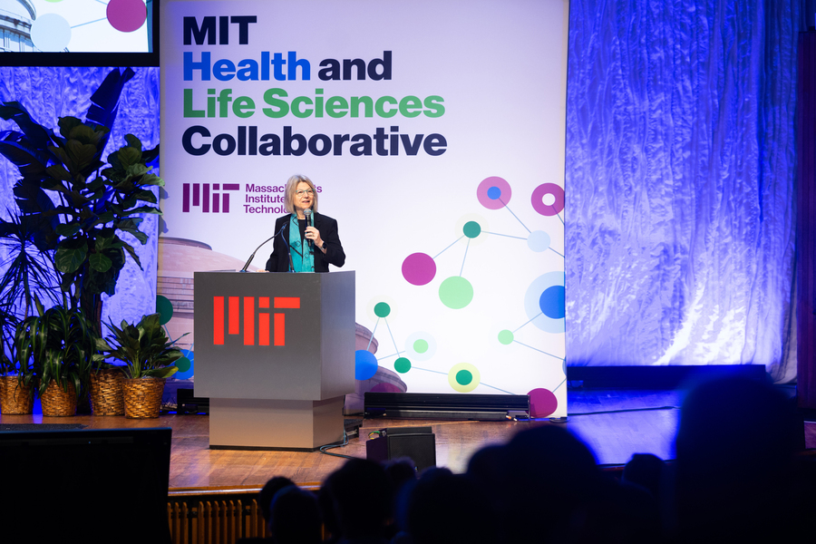
The MIT Health and Life Sciences Collaborative will bring together researchers from across the Institute to deliver health care solutions at scale.

Anne Trafton | MIT News
December 10, 2024

At MIT, collaboration between researchers working in the life sciences and engineering is a frequent occurrence. Under a new initiative launched last week, the Institute plans to strengthen and expand those collaborations to take on some of the most pressing health challenges facing the world.

The new MIT Health and Life Sciences Collaborative, or MIT HEALS, will bring together researchers from all over the Institute to find new solutions to challenges in health care. HEALS will draw on MIT’s strengths in life sciences and other fields, including artificial intelligence and chemical and biological engineering, to accelerate progress in improving patient care.

“As a source of new knowledge, of new tools and new cures, and of the innovators and the innovations that will shape the future of biomedicine and health care, there is just no place like MIT,” MIT President Sally Kornbluth said at a launch event last Wednesday in Kresge Auditorium. “Our goal with MIT HEALS is to help inspire, accelerate, and deliver solutions, at scale, to some of society’s most urgent and intractable health challenges.”

The launch event served as a day-long review of MIT’s historical impact in the life sciences and a preview of what it hopes to accomplish in the future.

“The talent assembled here has produced some truly towering accomplishments. But also — and, I believe, more importantly — you represent a deep well of creative potential for even greater impact,” Kornbluth said.

Massachusetts Governor Maura Healey, who addressed the filled auditorium, spoke of her excitement about the new initiative, emphasizing that “MIT’s leadership and the work that you do are more important than ever.”

“One of things as governor that I really appreciate is the opportunity to see so many of our state’s accomplished scientists and bright minds come together, work together, and forge a new commitment to improving human life,” Healey said. “It’s even more exciting when you think about this convening to think about all the amazing cures and treatments and discoveries that will result from it. I’m proud to say, and I really believe this, this is something that could only happen in Massachusetts. There’s no place that has the ecosystem that we have here, and we must fight hard to always protect that and to nurture that.”

A history of impact

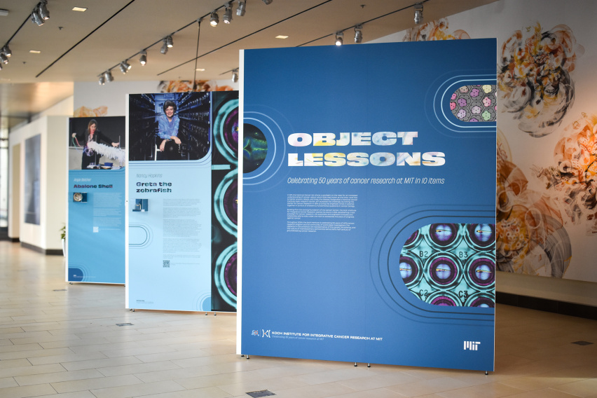
MIT has a long history of pioneering new fields in the life sciences, as MIT Institute Professor Phillip Sharp noted in his keynote address. Fifty years ago, MIT’s Center for Cancer Research was born, headed by Salvador Luria, a molecular biologist and a 1975 Nobel laureate.

That center helped to lead the revolutions in molecular biology, and later recombinant DNA technology, which have had significant impacts on human health. Research by MIT Professor Robert Weinberg and others identifying cancer genes has led the development of targeted drugs for cancer, including Herceptin and Gleevec.

In 2007, the Center for Cancer Research evolved into the Koch Institute for Integrative Cancer Research, whose faculty members are divided evenly between the School of Science and the School of Engineering, and where interdisciplinary collaboration is now the norm.

While MIT has long been a pioneer in this kind of collaborative health research, over the past several years, MIT’s visiting committees reported that there was potential to further enhance those collaborations, according to Nergis Mavalvala, dean of MIT’s School of Science.

“One of the very strong themes that emerged was that there’s an enormous hunger among our colleagues to collaborate more. And not just within their disciplines and within their departments, but across departmental boundaries, across school boundaries, and even with the hospitals and the biotech sector,” Mavalvala told MIT News.

To explore whether MIT could be doing more to encourage interdisciplinary research in the life sciences, Mavalvala and Anantha Chandrakasan, dean of the School of Engineering and MIT’s chief innovation and strategy officer, appointed a faculty committee called VITALS (Vision to Integrate, Translate and Advance Life Sciences).

That committee was co-chaired by Tyler Jacks, the David H. Koch Professor of Biology at MIT and a member and former director of the Koch Institute, and Kristala Jones Prather, head of MIT’s Department of Chemical Engineering.

“We surveyed the faculty, and for many people, the sense was that they could do more if there were improved mechanisms for interaction and collaboration. Not that those don’t exist — everybody knows that we have a highly collaborative environment at MIT, but that we could do even more if we had some additional infrastructure in place to facilitate bringing people together, and perhaps providing funding to initiate collaborative projects,” Jacks said before last week’s launch.

These efforts will build on and expand existing collaborative structures. MIT is already home to a number of institutes that promote collaboration across disciplines, including not only the Koch Institute but also the McGovern Institute for Brain Research, the Picower Institute for Learning and Memory, and the Institute for Medical Engineering and Science.

“We have some great examples of crosscutting work around MIT, but there’s still more opportunity to bring together faculty and researchers across the Institute,” Chandrakasan said before the launch event. “While there are these great individual pieces, we can amplify those while creating new collaborations.”

Supporting science

In her opening remarks on Wednesday, Kornbluth announced several new programs designed to support researchers in the life sciences and help promote connections between faculty at MIT, surrounding institutions and hospitals, and companies in the Kendall Square area.

“A crucial part of MIT HEALS will be finding ways to support, mentor, connect, and foster community for the very best minds, at every stage of their careers,” she said.

With funding provided by Noubar Afeyan PhD ’87, an executive member of the MIT Corporation and founder and CEO of Flagship Pioneering, MIT HEALS will offer fellowships for graduate students interested in exploring new directions in the life sciences.

Another key component of MIT HEALS will be the new Hood Pediatric Innovation Hub, which will focus on development of medical treatments specifically for children. This program, established with a gift from the Charles H. Hood Foundation, will be led by Elazer Edelman, a cardiologist and the Edward J. Poitras Professor in Medical Engineering and Science at MIT.

“Currently, the major market incentives are for medical innovations intended for adults — because that’s where the money is. As a result, children are all too often treated with medical devices and therapies that don’t meet their needs, because they’re simply scaled-down versions of the adult models,” Kornbluth said.

As another tool to help promising research projects get off the ground, MIT HEALS will include a grant program known as the MIT-MGB Seed Program. This program, which will fund joint research projects between MIT and Massachusetts General Hospital/Brigham and Women’s Hospital, is being launched with support from Analog Devices, to establish the Analog Devices, Inc. Fund for Health and Life Sciences.

Additionally, the Biswas Family Foundation is providing funding for postdoctoral fellows, who will receive four-year appointments to pursue collaborative health sciences research. The details of the fellows program will be announced in spring 2025.

“One of the things we have learned through experience is that when we do collaborative work that is cross-disciplinary, the people who are actually crossing disciplinary boundaries and going into multiple labs are students and postdocs,” Mavalvala said prior to the launch event. “The trainees, the younger generation, are much more nimble, moving between labs, learning new techniques and integrating new ideas.”

Revolutions

Discussions following the release of the VITALS committee report identified seven potential research areas where new research could have a big impact: AI and life science, low-cost diagnostics, neuroscience and mental health, environmental life science, food and agriculture, the future of public health and health care, and women’s health. However, Chandrakasan noted that research within HEALS will not be limited to those topics.

“We want this to be a very bottom-up process,” he told MIT News. “While there will be a few areas like AI and life sciences that we will absolutely prioritize, there will be plenty of room for us to be surprised on those innovative, forward-looking directions, and we hope to be surprised.”

At the launch event, faculty members from departments across MIT shared their work during panels that focused on the biosphere, brains, health care, immunology, entrepreneurship, artificial intelligence, translation, and collaboration. The program, which was developed by Amy Keating, head of the Department of Biology, and Katharina Ribbeck, the Andrew and Erna Viterbi Professor of Biological Engineering, also included a spoken-word performance by Victory Yinka-Banjo, an MIT senior majoring in computer science and molecular biology.

In her performance, called “Systems,” Yinka-Banjo urged the audience to “zoom out,” look at systems in their entirety, and pursue collective action.

“To be at MIT is to contribute to an era of infinite impact. It is to look beyond the microscope, zooming out to embrace the grander scope. To be at MIT is to latch onto hope so that in spite of a global pandemic, we fight and we cope. We fight with science and policy across clinics, academia, and industry for the betterment of our planet, for our rights, for our health,” she said.

In a panel titled “Revolutions,” Douglas Lauffenburger, the Ford Professor of Engineering and one of the founders of MIT’s Department of Biological Engineering, noted that engineers have been innovating in medicine since the 1950s, producing critical advances such as kidney dialysis, prosthetic limbs, and sophisticated medical imaging techniques.

MIT launched its program in biological engineering in 1998, and it became a full-fledged department in 2005. The department was founded based on the concept of developing new approaches to studying biology and developing potential treatments based on the new advances being made in molecular biology and genomics.

“Those two revolutions laid the foundation for a brand new kind of engineering that was not possible before them,” Lauffenburger said.

During that panel, Jacks and Ruth Lehmann, director of the Whitehead Institute for Biomedical Research, outlined several interdisciplinary projects underway at the Koch Institute and the Whitehead Institute. Those projects include using AI to analyze mammogram images and detect cancer earlier, engineering drought-resistant plants, and using CRISPR to identify genes involved in toxoplasmosis infection.

These examples illustrate the potential impact that can occur when “basic science meets translational science,” Lehmann said.

“I’m really looking forward to HEALS further enlarging the interactions that we have, and I think the possibilities for science, both at a mechanistic level and understanding the complexities of health and the planet, are really great,” she said.

The importance of teamwork

To bring together faculty and students with common interests and help spur new collaborations, HEALS plans to host workshops on different health-related topics. A faculty committee is now searching for a director for HEALS, who will coordinate these efforts.

Another important goal of the HEALS initiative, which was the focus of the day’s final panel discussion, is enhancing partnerships with Boston-area hospitals and biotech companies.

“There are many, many different forms of collaboration,” said Anne Klibanski, president and CEO of Mass General Brigham. “Part of it is the people. You bring the people together. Part of it is the ideas. But I have found certainly in our system, the way to get the best and the brightest people working together is to give them a problem to solve. You give them a problem to solve, and that’s where you get the energy, the passion, and the talent working together.”

Robert Langer, the David H. Koch Institute Professor at MIT and a member of the Koch Institute, noted the importance of tackling fundamental challenges without knowing exactly where they will lead. Langer, trained as a chemical engineer, began working in biomedical research in the 1970s, when most of his engineering classmates were going into jobs in the oil industry.

At the time, he worked with Judah Folkman at Boston Children’s Hospital on the idea of developing drugs that would starve tumors by cutting off their blood supply. “It took many, many years before those would [reach patients],” he says. “It took Genentech doing great work, building on some of the things we did that would lead to Avastin and many other drugs.”

Langer has spent much of his career developing novel strategies for delivering molecules, including messenger RNA, into cells. In 2010, he and Afeyan co-founded Moderna to further develop mRNA technology, which was eventually incorporated into mRNA vaccines for Covid.

“The important thing is to try to figure out what the applications are, which is a team effort,” Langer said. “Certainly when we published those papers in 1976, we had obviously no idea that messenger RNA would be important, that Covid would even exist. And so really it ends up being a team effort over the years.”

KI Gallery Exhibit: Artifacts from a half century of cancer research

Celebrating 50 years of MIT's cancer research program and the individuals who have shaped its journey, the Koch Institute Gallery features 10 significant artifacts, from one of the earliest PCR machine developed by Nobel Laureate H. Robert Horvitz to a preserved zebrafish from the lab of Nancy Hopkins in the Koch Institute Public Galleries. Visit Monday through Friday, 9AM-5PM.

Koch Institute
November 21, 2024

Throughout 2024, MIT’s Koch Institute for Integrative Cancer Research has celebrated 50 years of MIT’s cancer research program and the individuals who have shaped its journey. In honor of this milestone anniversary year, on November 19, the Koch Institute celebrated the opening of a new exhibition: Object Lessons: Celebrating 50 Years of Cancer Research at MIT in 10 Items. Object Lessons invites the public to explore significant artifacts—from one of the earliest PCR machines, developed in the lab of Nobel laureate H. Robert Horvitz, to Greta, a groundbreaking zebrafish from the lab of Professor Nancy Hopkins—in the half century of discoveries and advancements that have positioned MIT at the forefront of the fight against cancer.

50 years of innovation

The exhibition provides a glimpse into the many contributors and advancements that have defined MIT’s cancer research history since the founding of the Center for Cancer Research in 1974. When the National Cancer Act was passed in 1971, very little was understood about the biology of cancer, and it aimed to deepen our understanding of cancer and develop better strategies for the prevention, detection, and treatment of the disease. MIT embraced this call to action, establishing a center where many leading biologists tackled cancer’s fundamental questions. Building on this foundation, the Koch Institute opened its doors in 2011, housing engineers and life scientists from many fields under one roof to accelerate progress against cancer in novel and transformative ways.

In the 13 years since, the Koch Institute’s collaborative and interdisciplinary approach to cancer research has yielded significant advances in our understanding of the underlying biology of cancer and allowed for the translation of these discoveries into meaningful patient impacts. Over 120 spin-out companies—many headquartered nearby in the Kendall Square area—have their roots in Koch Institute research, with nearly half having advanced their technologies to clinical trials or commercial applications. The Koch Institute’s collaborative approach extends beyond its labs: principal investigators often form partnerships with colleagues at world-renowned medical centers, bridging the gap between discovery and clinical impact.

Current Koch Institute Director Matthew Vander Heiden, also a practicing oncologist at the Dana-Farber Cancer Institute, is driven by patient stories.

“It is never lost on us that the work we do in the lab is important to change the reality of cancer for patients,” he says. “We are constantly motivated by the urgent need to translate our research and improve outcomes for those impacted by cancer.”

Symbols of progress

The items on display as part of Object Lessons take viewers on a journey through five decades of MIT cancer research, from the pioneering days of Salvador Luria, founding director of the Center for Cancer Research, to some of the Koch Institute’s newest investigators including Francisco Sánchez-Rivera, Eisen and Chang Career Development Professor and an assistant professor of biology, and Jessica Stark, Underwood-Prescott Career Development Professor and an assistant professor of biological engineering and chemical engineering.

Among the standout pieces is a humble yet iconic object: Salvador Luria’s ceramic mug, emblazoned with “Luria’s broth.” Lysogeny broth, often called—apocryphally—Luria Broth, is a medium for growing bacteria. Still in use today, the recipe was first published in 1951 by a research associate in Luria’s lab. The artifact, on loan from the MIT Museum, symbolizes the foundational years of the Center for Cancer Research and serves as a reminder of Luria’s influence as an early visionary. His work set the stage for a new era of biological inquiry that would shape cancer research at MIT for generations.

Visitors can explore firsthand how the Koch Institute continues to build on the legacy of its predecessors, translating decades of knowledge into new tools and therapies that have the potential to transform patient care and cancer research.

For instance, the PCR machine designed in the Horvitz Lab in the 1980s made genetic manipulation of cells easier, and gene sequencing faster and more cost-effective. At the time of its commercialization, this groundbreaking benchtop unit marked a major leap forward. In the decades since, technological advances have allowed for the visualization of DNA and biological processes at a much smaller scale, as demonstrated by the handheld BioBits® imaging device developed by Stark and on display next door to the Horvitz panel.

 “We created BioBits kits to address a need for increased equity in STEM education,” Stark says. “By making hands-on biology education approachable and affordable, BioBits kits are helping inspire and empower the next generation of scientists.”

While the exhibition showcases scientific discoveries and marvels of engineering, it also aims to underscore the human element of cancer research through personally significant items, such as a messenger bag and Seq-Well device belonging to Alex Shalek, J. W. Kieckhefer Professor in the Institute for Medical Engineering and Science and the Department of Chemistry.

Shalek investigates the molecular differences between individual cells, developing mobile RNA-sequencing devices. He could often be seen toting the bag around the Boston area, and worldwide as he perfected and shared his technology with collaborators near and far. Through his work, Shalek has helped to make single cell sequencing accessible for labs in more than 30 countries across six continents.

“The KI seamlessly brings together students, staff, clinicians, and faculty across multiple different disciplines to collaboratively derive transformative insights into cancer,” Shalek says. “To me, these sorts of partnerships are the best part about being at MIT.”

Around the corner from Shalek’s display, visitors will find an object that serves as a stark reminder of the real people impacted by Koch Institute research: Steven Keating’s SM’12, PhD ’16 3D-printed model of his own brain tumor. Keating, who passed away in 2019, became a fierce advocate for the rights of patients to their medical data, and came to know Vander Heiden through his pursuit to become an expert on his tumor type, IDH-mutant glioma. In the years since, Vander Heiden’s work has contributed to a new therapy to treat Steven’s tumor type. In 2024, the drug, called vorasidenib, gained FDA approval, providing the first therapeutic breakthrough for Keating’s cancer in more than 20 years.

As the Koch Institute looks to the future, Object Lessons stands as a celebration of the people, the science, and the culture that have defined MIT’s first half-century of breakthroughs and contributions to the field of cancer research.

“Working in the uniquely collaborative environment of the Koch Institute and MIT, I am confident that we will continue to unlock key insights in the fight against cancer,” says Vander Heiden. “Our community is poised to embark on our next 50 years with the same passion and innovation that has carried us this far.”

Object Lessons will be on view in the Koch Institute Public Galleries. Visit Monday through Friday, 9 a.m. to 5 p.m., to see the exhibit up close.

Alumni Spotlight: Distillery Founder with a Spirited Passion

Jennifer Yang, '97, has been drawing on her biology degree for making spirits at a craft distillery in Maryland.

Jessica R. Simpson | Slice of MIT
October 15, 2024

If you had told Jennifer Yang ’97 during her time as a Course 7 major at MIT that she would use her biology degree to run a distillery, she wouldn’t have believed you.

“When I was at MIT, I looked at entrepreneurs and I thought, ‘Oh my gosh, that’s not me. I’m not one of those people who are so innovative and gutsy and brave,’” Yang says.

Managing a distillery is a passion that matured in Yang over time—much like the complex flavor of a barrel-aged whiskey. After graduating from MIT, the New York-native moved to Washington, DC, to pursue a career in management and technology consulting, which involved a lot of after-hours networking events. While building connections with colleagues over a glass of whiskey—a drink that was particularly popular with clients—Yang discovered her passion. Over the course of 10 years, she researched the science of making spirits, explored different small distilleries, and even started a whiskey tasting club.

“Being a science geek at heart and being very curious, I went down this rabbit hole pretty quickly in terms of wanting to learn more about it,” Yang explains.

In November 2022, she and her husband opened Covalent Spirits, a craft distillery, tasting room, and event space in Westminster, Maryland. In addition to producing bourbon whiskey, Covalent Spirits distills and blends vodka, gin, rum, and liqueurs. One of the bar’s unique and in-demand offerings is the “pH,” or “power of hydrogen,” cocktail, which uses the acidity of lemonade to turn a blue tea into a vibrant purple. Yang still works in consulting, but you can find her in her element behind the bar, engineering “pH” (and many other) cocktails Thursday through Saturday.

In her spare time, Yang is a committed MIT volunteer. An active participant in the Club of Washington DC, she is the regional alumni ambassador for the Baltimore area as well. Yang is also an educational counselor and the current president of the Class of 1997. She notes that she and her ’97 classmates were the first to organize pi reunions, a tradition in which alums gather in Las Vegas 3.14 years after graduation. “We’re glad our class could leave a little bit of a legacy,” she says.

In fact, the shared MIT connection between alumni inspired Yang to name her company Covalent Spirits. One year, at an MIT gathering, Yang started talking to another alum about planning events for undergrad classes that shared years at MIT—what they called “covalent classes.” Yang has since incorporated literal and metaphorical covalent bonds (a chemical connection between atoms formed by sharing) into every facet of her business: from the chemistry of making spirits, to the design of the distillery logo, to the company’s emphasis on community.

“While we are striving to create really good products, we also want to create a space and experiences for people to get together and geek out over a common interest, to celebrate an occasion, or to connect over anything,” Yang elaborates. “You share a drink, you share an experience, you share a community. Bonding through sharing is the covalent spirit.”

A new approach to modeling complex biological systems

MIT engineers’ new model could help researchers glean insights from genomic data and other huge datasets. This is potentially critical to researchers who study any kind of complex biological system, according to senior author Douglas Lauffenburger.

Anne Trafton | MIT News
November 5, 2024

Over the past two decades, new technologies have helped scientists generate a vast amount of biological data. Large-scale experiments in genomics, transcriptomics, proteomics, and cytometry can produce enormous quantities of data from a given cellular or multicellular system.

However, making sense of this information is not always easy. This is especially true when trying to analyze complex systems such as the cascade of interactions that occur when the immune system encounters a foreign pathogen.

MIT biological engineers have now developed a new computational method for extracting useful information from these datasets. Using their new technique, they showed that they could unravel a series of interactions that determine how the immune system responds to tuberculosis vaccination and subsequent infection.

This strategy could be useful to vaccine developers and to researchers who study any kind of complex biological system, says Douglas Lauffenburger, the Ford Professor of Engineering in the departments of Biological Engineering, Biology, and Chemical Engineering.

“We’ve landed on a computational modeling framework that allows prediction of effects of perturbations in a highly complex system, including multiple scales and many different types of components,” says Lauffenburger, the senior author of the new study.

Shu Wang, a former MIT postdoc who is now an assistant professor at the University of Toronto, and Amy Myers, a research manager in the lab of University of Pittsburgh School of Medicine Professor JoAnne Flynn, are the lead authors of a new paper on the work, which appears today in the journal Cell Systems.

Modeling complex systems

When studying complex biological systems such as the immune system, scientists can extract many different types of data. Sequencing cell genomes tells them which gene variants a cell carries, while analyzing messenger RNA transcripts tells them which genes are being expressed in a given cell. Using proteomics, researchers can measure the proteins found in a cell or biological system, and cytometry allows them to quantify a myriad of cell types present.

Using computational approaches such as machine learning, scientists can use this data to train models to predict a specific output based on a given set of inputs — for example, whether a vaccine will generate a robust immune response. However, that type of modeling doesn’t reveal anything about the steps that happen in between the input and the output.

“That AI approach can be really useful for clinical medical purposes, but it’s not very useful for understanding biology, because usually you’re interested in everything that’s happening between the inputs and outputs,” Lauffenburger says. “What are the mechanisms that actually generate outputs from inputs?”

To create models that can identify the inner workings of complex biological systems, the researchers turned to a type of model known as a probabilistic graphical network. These models represent each measured variable as a node, generating maps of how each node is connected to the others.

Probabilistic graphical networks are often used for applications such as speech recognition and computer vision, but they have not been widely used in biology.

Lauffenburger’s lab has previously used this type of model to analyze intracellular signaling pathways, which required analyzing just one kind of data. To adapt this approach to analyze many datasets at once, the researchers applied a mathematical technique that can filter out any correlations between variables that are not directly affecting each other. This technique, known as graphical lasso, is an adaptation of the method often used in machine learning models to strip away results that are likely due to noise.

“With correlation-based network models generally, one of the problems that can arise is that everything seems to be influenced by everything else, so you have to figure out how to strip down to the most essential interactions,” Lauffenburger says. “Using probabilistic graphical network frameworks, one can really boil down to the things that are most likely to be direct and throw out the things that are most likely to be indirect.”

Mechanism of vaccination

To test their modeling approach, the researchers used data from studies of a tuberculosis vaccine. This vaccine, known as BCG, is an attenuated form of Mycobacterium bovis. It is used in many countries where TB is common but isn’t always effective, and its protection can weaken over time.

In hopes of developing more effective TB protection, researchers have been testing whether delivering the BCG vaccine intravenously or by inhalation might provoke a better immune response than injecting it. Those studies, performed in animals, found that the vaccine did work much better when given intravenously. In the MIT study, Lauffenburger and his colleagues attempted to discover the mechanism behind this success.

The data that the researchers examined in this study included measurements of about 200 variables, including levels of cytokines, antibodies, and different types of immune cells, from about 30 animals.

The measurements were taken before vaccination, after vaccination, and after TB infection. By analyzing the data using their new modeling approach, the MIT team was able to determine the steps needed to generate a strong immune response. They showed that the vaccine stimulates a subset of T cells, which produce a cytokine that activates a set of B cells that generate antibodies targeting the bacterium.

“Almost like a roadmap or a subway map, you could find what were really the most important paths. Even though a lot of other things in the immune system were changing one way or another, they were really off the critical path and didn’t matter so much,” Lauffenburger says.

The researchers then used the model to make predictions for how a specific disruption, such as suppressing a subset of immune cells, would affect the system. The model predicted that if B cells were nearly eliminated, there would be little impact on the vaccine response, and experiments showed that prediction was correct.

This modeling approach could be used by vaccine developers to predict the effect their vaccines may have, and to make tweaks that would improve them before testing them in humans. Lauffenburger’s lab is now using the model to study the mechanism of a malaria vaccine that has been given to children in Kenya, Ghana, and Malawi over the past few years.

“The advantage of this computational approach is that it filters out many biological targets that only indirectly influence the outcome and identifies those that directly regulate the response. Then it’s possible to predict how therapeutically altering those biological targets would change the response. This is significant because it provides the basis for future vaccine and trial designs that are more data driven,” says Kathryn Miller-Jensen, a professor of biomedical engineering at Yale University, who was not involved in the study.

Lauffenburger’s lab is also using this type of modeling to study the tumor microenvironment, which contains many types of immune cells and cancerous cells, in hopes of predicting how tumors might respond to different kinds of treatment.

The research was funded by the National Institute of Allergy and Infectious Diseases.

Research Reflections: Alison Biester (PhD ’24), Drennan Lab

New snapshots of ancient life

Alison Biester | Department of Chemistry
October 3, 2024

The resolution revolution, beating “blobology”, and shedding light on how ancient microbes thrived in a primordial soup.

The earliest life on earth created biological molecules despite the limited materials available in the primordial soup such as CO2, hydrogen gas, and minerals containing iron, nickel, and sulfur.

As ancient microbes evolved, they developed proteins that sped up chemical reactions, called enzymes. Enzymes were evolutionarily advantageous because they created local environments called active sites optimized for reaction performance.

Although we know that carbon is the building block of life on earth–we wouldn’t exist without carbon-based molecules such as proteins and DNA–much remains unclear about how more complex carbon-based molecules were originally generated from CO2. Proteins and DNA are huge molecules with thousands of carbon atoms, so creating life from CO2 would be no small undertaking.

Catherine Drennan, Professor of Biology and Chemistry and HHMI Investigator and Professor, has long studied the enzymes that perform these crucial reactions wherein CO2 is converted into a form of carbon that cells can use, which requires iron, nickel, and sulfur.

In particular, she uses structural biology to study carbon monoxide dehydrogenase (CODH), which reacts with CO2 to produce CO, and acetyl-CoA synthase (ACS), which uses CO with another single unit of carbon to create a carbon-carbon bond. Crystallographic work by Drennan and others has provided structural snapshots of bacterial CODH and ACS, but its structure in other contexts remains elusive. During my PhD, I worked with Drennan on the structural characterization of CODH and ACS, culminating in a publication in PNAS, published October 3, 2024.

Throughout Drennan’s career, the lab has used a method known as X-ray crystallography to determine enzyme structures at atomic resolution. In recent years, however, cryogenic electron microscopy (cryo-EM) has risen in popularity as a structural biology technique.

Cryo-EM offers some key advantages over X-ray crystallography, such as its ability to capture structures of large and dynamic complexes. However, cryo-EM is limited in its ability to elucidate structures of small proteins, an area where X-ray crystallography continues to excel.

To perform a cryo-EM experiment, proteins are rapidly frozen in a thin layer of ice and imaged on an electron microscope. By capturing images of the protein in various orientations, researchers can generate a 3D model of their protein of interest.

Around 2015, cryo-EM reached a tipping point known as the “resolution revolution.” Due to improvements in both the hardware for collecting cryo-EM data and the software used for data processing, the technique could, for the first time, be used to determine protein structures at near-atomic resolution.

Seeing the potential for this new technique, MIT opened its very own cryo-EM facility with two electron microscopes in 2018. Just a year later, I joined the Drennan lab. When I began my thesis work, Cathy asked “Would you like to do crystallography or cryo-EM?”

Eager to try something that was both novel for researchers and new to me, I chose cryo-EM.

Ancient microbes

An ancient type of microbe, archaea, also uses CODH and ACS. Without information on how these protein chains interact, we cannot understand how these proteins work together within this complex–but it’s a difficult question to answer. In total, the complex contains forty protein chains that interact with one another and adopt various conformations to perform their chemistry.

We don’t know for sure which ACS enzyme came first, the bacterial or archaeal one, but we know they are both very ancient.

Archaeal CODH has been visualized via X-ray crystallography, but that CODH was isolated from the enormous megadalton enzyme complex present in the native archaea.

A CO2 molecule, which reacts with CODH, is 44 daltons; the enzyme complex at 2.2 megadaltons is 50,000 times the size of CO2. The complex consists of several copies of CODH, ACS, and a cobalt-containing enzyme that donates the second one-carbon unit used by ACS. Due to the large and dynamic nature of the complex, it was a great candidate for visualizing with cryo-EM.

Before I joined the lab, a collaboration had been initiated between the Drennan Lab and Dr. David Grahame of the Uniformed Services University of the Health Sciences, an expert in archaeal CODH and ACS.

Just before his retirement, Grahame grew hundreds of liters of archaea and isolated approximately one gram of the enzyme complex that he provided to the Drennan Lab for structural characterization. Each cryo-EM experiment can use as little as a microgram of protein. For a structural biologist, having one gram of protein–in theory, enough for one million experiments–to work with is a dream.

Blobology

With an abundance of protein, I embarked on this project with this exciting new technique on a promising target. I prepared my cryo-EM sample and collected data at the new MIT cryo-EM facility. As I was collecting data, I could see in the images large protein complexes that appeared to be my complex of interest. I could also see some smaller proteins that were consistent with the shape of isolated CODH. When I went on to process my data, I focused on the larger protein complexes, since the structure of isolated CODH was already known.

However, when I finished processing my first dataset, I was a bit disappointed. My resolution was very low–instead of atoms, I was seeing amorphous blobs, and I had no idea which blob matched with which protein, or how the proteins fit together. Rather than post-revolution cryo-EM, I felt like I was performing the “blobology” of the past.

Our cryo-EM data contains detailed structural information that becomes evident after significant data processing. On the left is the initial structure of our proteins of interest, carbon monoxide dehydrogenase (CODH) and acetyl-CoA synthase (ACS), and on the right is our final, detailed one. Photo courtesy of Alison Biester.

But the project was young, and a few failed experiments are par for the course of a PhD.

The next step was sample optimization, and luckily I had plenty of sample to work with. I tried preparing the protein in a different way, changed the protein concentration, used different additives, and scaled up my data collection.

Nothing helped. No matter what I tried, I could not move out of blobology territory. So, as one does when a project is failing, I stepped away. I worked on other projects and stopped thinking about the archaeal CODH and ACS.

A few months later, the cryo-EM facility was seeking users to try a new sample preparation instrument called the chameleon. Chameleon automates the sample preparation process and is intended to improve sample quality. With plenty of sample still to spare, I volunteered to try the instrument.

Just prior to my data collection, the facility had also installed a new software that allows data processing as it is being collected. The software uses automated processes to select proteins within your data; previously, I had only selected large protein complexes consistent with my complex of interest after the fact.

The new software is not very discriminating–but I was surprised when I looked at the results of the live processing. The processing showed that I had a protein complex in my sample that I did not expect – a complex of CODH and ACS!

This complex had just one copy of CODH and one copy of ACS, unlike the full complex that has multiple copies of each. My excitement for the project was reinvigorated. With this new target, could I leave blobology behind and finally join the resolution revolution?

After running more experiments and collecting more data and a few months of data processing, I realized that the sample contained three different states: isolated CODH, CODH with one copy of ACS, and CODH with two copies of ACS. I was able to use the Model-based Analysis of Volume Ensembles (MAVEn) tool developed by the Davis Lab at MIT to sort out these three states. When I finished the data processing, I achieved near-atomic resolution of all three states.

Through this work, for the first time, we can see what the archaeal ACS looks like. The archaeal ACS is fundamentally different from the bacterial one: a huge portion of the enzyme is missing, including part of the enzyme that makes up the active site in bacteria, leaving open the question of what the ACS active site looks like in archaea.

In our structure of archaeal ACS in complex with CODH, we were surprised to see that the active site looks almost identical to the bacterial one. This similarity is enabled by the archaeal CODH, which compensates for the missing part of ACS.

Given how similar the ACS active site environment in bacterial and archaea, we are likely getting a look at an active site that has remained conserved over billions of years of evolution.

Although the project didn’t fulfill its original promise of solving the structure of the large, dynamic protein complex, I did find intriguing insights. The tools available in 2015 would not have enabled me to achieve these results; it is clear to me that the resolution revolution is far from over, and the evolution of structural biology has been fascinating to experience. Cryo-EM has and will continue to evolve, as amazing new tools are still being developed.

Since graduating from MIT, I’ve been working at the Protein Data Bank, the data center that houses all available protein structure information. Working here gives me a front-row view of new discoveries in structural biology. I’m so excited to see where this field will go in the future.

BSG-MSRP-Bio Student Profile: Adriana Camacho-Badillow, Calo Lab

Understanding the Role of PARPs and UBF1 in Building Ribosomes

Noah Daly | Department of Biology
September 25, 2024

While pursuing her passion for research, BSG-MSRP-Bio student Adriana Camacho-Badillo made major contributions to research in the Calo Lab in the Department of Biology at MIT.

Growing up in Puerto Rico, Adriana Camacho-Badillo had no explanation for her recurrent multiple fracture injuries. In her teens, she was finally able to see a geneticist who diagnosed her with a genetic syndrome that affects connective tissue throughout the body. 

This awakened an interest in genetics that led her to immerse herself in her genetic panel results, curious about the role of each gene that was tested. 

“I realized I wanted to find out how mutations affect gene expression that could possibly lead to a distinct phenotype or even a genetic syndrome,” she says. 

Within a few years of setting her sights on becoming a scientist, Camacho-Badillo began her first research experience working in the laboratory of Professors Hector Areizaga-Martínez and Elddie Román-Morales. Her work focused on experiments using enzymes to degrade Dichloro-diphenyl-trichloroethane, or DDT, a once-common pesticide known to be highly toxic to humans and other mammals that remains in the environment long after application to crops. 

As she became familiar with the day-to-day routines of designing and executing research experiments, she realized she was drawn to biochemistry and molecular biology. Camacho-Badillo soon applied to the molecular neuroscience lab of Professor Miguel Méndez at the University of Puerto Rico at Aguadilla and joined their team working on the effects of high glucose in the central nervous system of mice.

Expanding Experiences While Narrowing Focus

When Camacho-Badillo was sixteen, alongside Méndez and other students, she participated in the Quantitative Methods Workshop at MIT. The workshop allows undergraduate students from universities around the United States and the Caribbean to come together for a few days in January to learn how to apply computational tools that can help biological research. 

One of the sessions she attended was a talk about machine learning and studying the brain, presented by graduate student Taylor Baum. 

“I loved Taylor’s workshop,” Camacho-Badillo said, “When Taylor asked if anyone would be interested in volunteering to teach Spanish-speaking students in grade school science, I said yes without hesitation.” 

Baum, a neuroscientist and computer scientist working in the Munther Dahleh Research Group at MIT, is also the founder of Sprouting, Inc. The organization equips high-school students and undergraduates in Puerto Rico with STEM skills to help them pursue careers in science and technology.

After participating in QMW, it wasn’t long before Camacho-Badillo was back at MIT. She participated in the Bernard S. and Sophie G. Gould MIT Summer Research Program in Biology in 2023 and worked in the Yamashita Lab, studying two phenotypes of genetic mutations associated with cancer during cell division. 

The BSG-MSRP-Bio program offers lab experience and extracurricular activities such as journal clubs and dinners with professors. At one of these events, she met Associate Professor of Biology Eliezer Calo.

Camacho-Badillo and her mentor Eliezer Calo, Associate Professor of Biology. Photo Credit: Mandana Sassanfar.

“I loved meeting another scientist from Puerto Rico working on molecular biology, so I decided to look further into his research,” Camacho-Badillo recalls. 

In 2024, she was delighted to have the opportunity to return to the BSG-MSRP-Bio Program for a second time, and now to work in Calo’s Lab. 

The Unsolved Mysteries of UBF1

Although BSG-MSRP-Bio students are often mentored by graduate students or postdocs, Calo spent the summer mentoring Camacho-Badillo directly. As an alumnus of the MSRP-Bio program himself, Calo understands firsthand how much of an impact meaningful research can have for an undergraduate student spending a few months experiencing life in the lab at MIT. 

In the Calo Lab, Camacho-Badillo spent the early days of this summer poring over past research papers on genetic transcription, trying to answer a big question in molecular biology. Camacho-Badillo has been helping Calo understand how a particular protein affects the production of ribosomes in cells.

A ribosome is the molecular machinery that synthesizes proteins, and an average cell can produce around 10 million ribosomes to sustain its essential functions. Creating these protein engines requires the transcription of ribosomal DNA, or rDNA. 

In order to synthesize RNA, specific proteins called polymerases must bind to the DNA. Camacho-Badillo’s work focuses on one of those binding proteins called upstream binding factor, or UBF1. UBF1 is essential for the synthesis of the ribosomal RNA. The UBF1 transcription factor is responsible for recruiting the polymerase, RNA polymerase I, to transcribe the rDNA into rRNA.

Despite knowing the importance of UBF1 in ribosomal production, it’s unclear what its full purpose is in this process. Calo and Camacho-Badillo think that clarifying the role of UBF1 in ribosomal biogenesis will help scientists understand how certain neurological diseases occur. UBF1 is known to be associated with diseases such as acute myeloid leukemia and childhood-onset neurodegeneration with brain atrophy, but the mechanism is not yet understood.

UBF1 is a peculiar transcription factor. Before it can transcribe a gene, UBF1 must first dimerize, forming a bond with another UBF1 protein. After binding to the rDNA, UBF1 can recruit the remaining RNA transcription machinery. The dimer is crucial for transcription to occur, yet this protein can make further connections with other UBF1 monomers, a process called oligomerization. 

Nothing is concretely understood about how oligomers of UBF1 form: they could be critical for transcription, forming clusters that can no longer bind with rDNA or inhibit the recruitment of the remaining RNA transcription machinery. These clusters could also be directly contributing to a variety of neurological diseases.

“The genome contains multiple rDNA copies, but not all are utilized,” Calo explains. “UBF1 must precisely identify the correct copies to activate while avoiding the formation of aggregates that could impair its function.”

The regulation of these dimers is also a mystery. Early in the summer, Camacho-Badillo helped make an important connection: prior research from the Calo Lab showed that enzymes called poly ADP-ribose polymerases, or PARPs, play a role in maintaining chemical properties in the nucleolus, where ribosomes are produced and assembled. The main target of these proteins within the RNA transcriptional machinery before transcription is initiated is UBF1.  

Based on this initial result, Camacho-Badillo’s entire summer project shifted to further characterize PARPs in ribosome biogenesis.

“This observation about the role PARPs plays is a big deal for us,” Calo says. “We do many experiments in my lab, but Adriana’s work this summer has opened a key gateway to understanding the mysteries behind UBF1 regulation, leading to proper ribosome production and allowing the Calo lab to pursue this goal. She’s going to be a superstar.” 

Camacho-Badillo’s work hasn’t ended with the BSG-MSRP-Bio program, however. She’ll spend the fall semester at MIT, continuing to work on understanding how rDNA transcription is regulated as a visiting student in the Calo Lab. Although she still has a year and a half to go in her undergraduate degree, she’s already set her sights on graduate school. 

“This program has meant so much to me and brought so much into my life,” she says. “All I want to do right now is keep this research going.”

Want to know more about our BSG-MSRP-Bio Students? Read more testimonials and stories here.

From open education learners to MIT coders

MIT Digital Learning Lab’s high school interns gain professional experience working on the backend of open online MITx courses. The program emerged after Mary Ellen Wiltrout, PhD '09, digital learning scientist at MIT Open Learning, connected with the executive director and founder of Empowr, a nonprofit that serves low-income communities by creating a school-to-career pipeline through software development skills.

Katherine Ouellette | MIT Open Learning
August 26, 2024

Switching programming languages is not as simple as switching word processors. Yet high schooler Thomas Esayas quickly adapted from Swift to Python during his 2023 internship with the MIT Digital Learning Laba joint program between MIT Open Learning and the Institute’s academic departments. One year later, Esayas returns to the Institute for a second internship and as a new undergraduate student.

“I felt thoroughly challenged and learned a lot of new skills,” says Esayas.

Through this remote opportunity, interns gain real-world coding experience and practice professional skills by collaborating on MIT’s open online courses. The four interns from Digital Learning Lab’s 2023 and 2024 cohorts also participate in Empowr, a four-year program for low-income high school students that teaches in-demand software development skills and helps them secure paid internships.

The Digital Learning Lab program emerged after Mary Ellen Wiltrout PhD ’09, digital learning scientist at MIT Open Learning, connected with Adrian Devezin, executive director and founder of Empowr, at a conference about making education more accessible and equitable.

“It was affirming to have someone else see what Empowr is trying to do,” says Devezin about the organization’s goal to strengthen the school-to-career pipeline. “Being able to collaborate was beautiful for me, and more importantly, to the students.”

Building technical skills and self-confidence

The Digital Learning Lab internship empowers students to build confidence in their technical abilities, career skills, and the college application process. Interns assist the lab’s digital learning scientists with their work developing and maintaining online MITx courses at Open Learning across multiple academic areas.

“I found myself always busy with something interesting to work on,” says Esayas.

The interactive open education resources that Esayas produced last summer are now being used in live courses. He also helped find and fix bugs on the platform that hosts the MITx courses.

The internship’s flexible design allows projects to be adapted based on the student’s personal progress and interests.

“The students became co-creators of their educational experiences,” says Wiltrout, noting this is beneficial from a pedagogical standpoint.

Devezin adds, “I definitely saw a big improvement in their problem-solving abilities. Having to switch their mindset to a new language, work in new frameworks, and work on teams solving real problems enhanced their ability to adapt to new situations.”

The students’ also strengthened their professional repertoire in areas such as collaboration, communication, and project management. The 2023 cohort, Devezin says, developed the initiative to help other students and take on leadership roles.

Now that Esayas has completed his 2024 internship, he says, “I’m glad that I got to collaborate with more people and work on more projects. Overall, I’m very happy I was able to return.”

two people smiling, standing in front of a colorful wall.
Adrian Devezin, executive director and founder of Empowr (left), and Mary Ellen Wiltrout, digital learning scientist at MIT Open Learning (right), presented their takeaways from the first year of the MIT Digital Learning Lab internship at the 2024 Open edX conference. Photo courtesy of Empowr.

Learning from both sides

Learning occurred for both students and educators alike. Wiltrout says that the Digital Learning Lab values the opportunity to see the interns’ growth day-to-day and week-to-week, since digital learning scientists rarely follow the trajectory of individual learners who are using the course materials they create. Having instant feedback informs how they can adjust their teaching approaches for various problems.

The positive impact of the Digital Learning Lab internship’s hands-on learning experiences has made Devezin rethink the way he teaches class moving forward, and “the problems I want them to be solving,” he says.

Now, Devezin tries to emulate the real-world experience of working on a project for his Empowr students. Instead of assigning coding exercises where he provides the exact methods to solve the problems, he started asking students to determine the correct approach on their own.

The fact that Wiltrout and Devezin are open to adapting their teaching methods based on student feedback is indicative of a key factor to the internship’s success — active participation in students’ growth. It was mutually beneficial for the students and the educators to have determined stakeholders at both Digital Learning Lab and Empowr.

“A lot of dedicated educators understand that there’s a lot of inequities in education, and we need to come together to solve them,” Devezin says.

The Digital Learning Lab internship shows how open source learning materials can make educational and professional opportunities more accessible. The 2024 cohort has been able to increase their annual household income by an average of 75%, a recent Empowr report revealed. Wiltrout says that the two new Empowr students seem more confident with coding and showed enthusiasm and dedication to their tasks as they also consider colleges.

Wiltrout and Devezin presented their takeaways from the internship’s first year at the 2024 Open edX conference.

“I think it’s important to try making sure that more people are aware of tools and resources that are out there,” Wiltrout says. “Then giving people opportunities where they may not have otherwise had that chance.”

Now, Devezin is thinking about how Empowr students can come full circle with their relationship to open educational materials. He’s asking, “How can I help my students contribute to the open source world to give back to others?”