A sum of their parts

Researchers in the Department of Biology at MIT use an AI-driven approach to computationally predict short amino acid sequences that can bind to or inhibit a target, with a potential for great impact on fundamental biological research and therapeutic applications.

Lillian Eden | Department of Biology
February 6, 2025

All biological function is dependent on how different proteins interact with each other. Protein-protein interactions facilitate everything from transcribing DNA and controlling cell division to higher-level functions in complex organisms.

Much remains unclear about how these functions are orchestrated on the molecular level, however, and how proteins interact with each other — either with other proteins or with copies of themselves. 

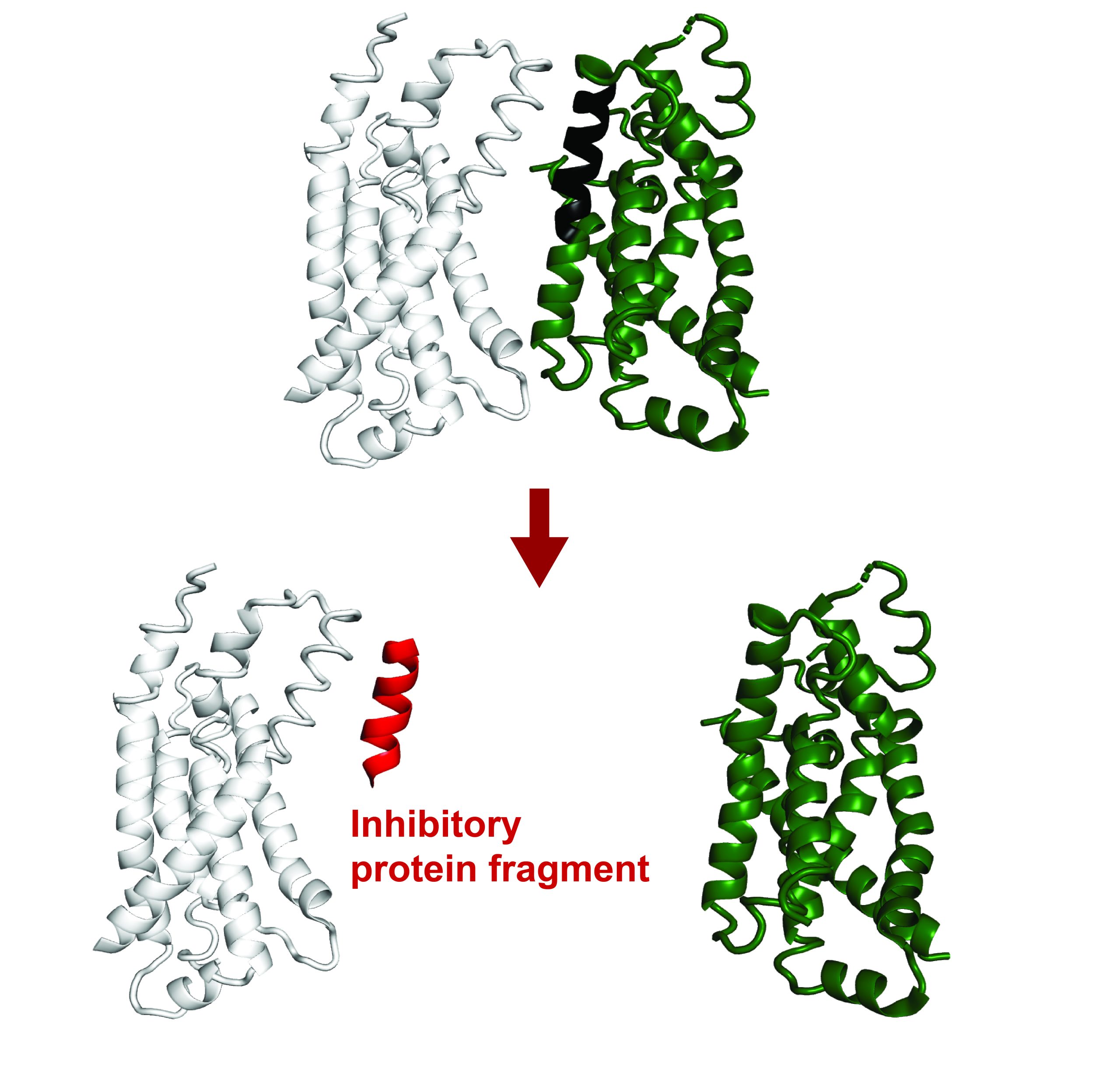
Recent findings have revealed that small protein fragments have a lot of functional potential. Even though they are incomplete pieces, short stretches of amino acids can still bind to interfaces of a target protein, recapitulating native interactions. Through this process, they can alter that protein’s function or disrupt its interactions with other proteins. 

Protein fragments could therefore empower both basic research on protein interactions and cellular processes and could potentially have therapeutic applications. 

Recently published in Proceedings of the National Academy of Sciences, a new computational method developed in the Department of Biology at MIT builds on existing AI models to computationally predict protein fragments that can bind to and inhibit full-length proteins in E. coli. Theoretically, this tool could lead to genetically encodable inhibitors against any protein. 

The work was done in the lab of Associate Professor of Biology and HHMI Investigator Gene-Wei Li in collaboration with the lab of Jay A. Stein (1968) Professor of Biology, Professor of Biological Engineering and Department Head Amy Keating.

Leveraging Machine Learning

The program, called FragFold, leverages AlphaFold, an AI model that has led to phenomenal advancements in biology in recent years due to its ability to predict protein folding and protein interactions. 

The goal of the project was to predict fragment inhibitors, which is a novel application of AlphaFold. The researchers on this project confirmed experimentally that more than half of FragFold’s predictions for binding or inhibition were accurate, even when researchers had no previous structural data on the mechanisms of those interactions. 

“Our results suggest that this is a generalizable approach to find binding modes that are likely to inhibit protein function, including for novel protein targets, and you can use these predictions as a starting point for further experiments,” says co-first and corresponding author Andrew Savinov, a postdoc in the Li Lab. “We can really apply this to proteins without known functions, without known interactions, without even known structures, and we can put some credence in these models we’re developing.”

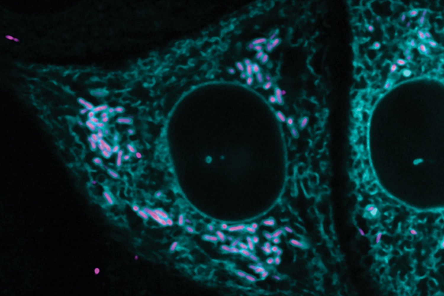
One example is FtsZ, a protein that is key for cell division. It is well-studied but contains a region that is intrinsically disordered and, therefore, especially challenging to study. Disordered proteins are dynamic, and their functional interactions are very likely fleeting — occurring so briefly that current structural biology tools can’t capture a single structure or interaction. 

The researchers leveraged FragFold to explore the activity of fragments of FtsZ, including fragments of the intrinsically disordered region, to identify several new binding interactions with various proteins. This leap in understanding confirms and expands upon previous experiments measuring FtsZ’s biological activity. 

This progress is significant in part because it was made without solving the disordered region’s structure, and because it exhibits the potential power of FragFold.

“This is one example of how AlphaFold is fundamentally changing how we can study molecular and cell biology,” Keating says. “Creative applications of AI methods, such as our work on FragFold, open up unexpected capabilities and new research directions.”

Inhibition, and beyond

The researchers accomplished these predictions by computationally fragmenting each protein and then modeling how those fragments would bind to interaction partners they thought were relevant.

They compared the maps of predicted binding across the entire sequence to the effects of those same fragments in living cells, determined using high-throughput experimental measurements in which millions of cells each produce one type of protein fragment. 

AlphaFold uses co-evolutionary information to predict folding, and typically evaluates the evolutionary history of proteins using something called multiple sequence alignments for every single prediction run. The MSAs are critical, but are a bottleneck for large-scale predictions — they can take a prohibitive amount of time and computational power. 

For FragFold, the researchers instead pre-calculated the MSA for a full-length protein once and used that result to guide the predictions for each fragment of that full-length protein. 

Savinov, together with Keating Lab alum Sebastian Swanson, PhD ‘23, predicted inhibitory fragments of a diverse set of proteins in addition to FtsZ. Among the interactions they explored was a complex between lipopolysaccharide transport proteins LptF and LptG. A protein fragment of LptG inhibited this interaction, presumably disrupting the delivery of lipopolysaccharide, which is a crucial component of the E. coli outer cell membrane essential for cellular fitness.

“The big surprise was that we can predict binding with such high accuracy and, in fact, often predict binding that corresponds to inhibition,” Savinov says. “For every protein we’ve looked at, we’ve been able to find inhibitors.”

The researchers initially focused on protein fragments as inhibitors because whether a fragment could block an essential function in cells is a relatively simple outcome to measure systematically. Looking forward, Savinov is also interested in exploring fragment function outside inhibition, such as fragments that can stabilize the protein they bind to, enhance or alter its function, or trigger protein degradation. 

Design, in principle 

This research is a starting point for developing a systemic understanding of cellular design principles, and what elements deep-learning models may be drawing on to make accurate predictions. 

“There’s a broader, further-reaching goal that we’re building towards,” Savinov says. “Now that we can predict them, can we use the data we have from predictions and experiments to pull out the salient features to figure out what AlphaFold has actually learned about what makes a good inhibitor?” 

Savinov and collaborators also delved further into how protein fragments bind, exploring other protein interactions and mutating specific residues to see how those interactions change how the fragment interacts with its target. 

Experimentally examining the behavior of thousands of mutated fragments within cells, an approach known as deep mutational scanning, revealed key amino acids that are responsible for inhibition. In some cases, the mutated fragments were even more potent inhibitors than their natural, full-length sequences. 

“Unlike previous methods, we are not limited to identifying fragments in experimental structural data,” says Swanson. “The core strength of this work is the interplay between high-throughput experimental inhibition data and the predicted structural models: the experimental data guides us towards the fragments that are particularly interesting, while the structural models predicted by FragFold provide a specific, testable hypothesis for how the fragments function on a molecular level.”

Savinov is excited about the future of this approach and its myriad applications.

“By creating compact, genetically encodable binders, FragFold opens a wide range of possibilities to manipulate protein function,” Li agrees. “We can imagine delivering functionalized fragments that can modify native proteins, change their subcellular localization, and even reprogram them to create new tools for studying cell biology and treating diseases.” 

Kingdoms collide as bacteria and cells form captivating connections

Studying the pathogen R. parkeri, researchers discovered the first evidence of extensive and stable interkingdom contacts between a pathogen and a eukaryotic organelle.

Lillian Eden | Department of Biology
January 24, 2025

In biology textbooks, the endoplasmic reticulum is often portrayed as a distinct, compact organelle near the nucleus, and is commonly known to be responsible for protein trafficking and secretion. In reality, the ER is vast and dynamic, spread throughout the cell and able to establish contact and communication with and between other organelles. These membrane contacts regulate processes as diverse as fat metabolism, sugar metabolism, and immune responses.

Exploring how pathogens manipulate and hijack essential processes to promote their own life cycles can reveal much about fundamental cellular functions and provide insight into viable treatment options for understudied pathogens.

New research from the Lamason Lab in the Department of Biology at MIT recently published in the Journal of Cell Biology has shown that Rickettsia parkeri, a bacterial pathogen that lives freely in the cytosol, can interact in an extensive and stable way with the rough endoplasmic reticulum, forming previously unseen contacts with the organelle.

It’s the first known example of a direct interkingdom contact site between an intracellular bacterial pathogen and a eukaryotic membrane.

The Lamason Lab studies R. parkeri as a model for infection of the more virulent Rickettsia rickettsii. R. rickettsii, carried and transmitted by ticks, causes Rocky Mountain Spotted Fever. Left untreated, the infection can cause symptoms as severe as organ failure and death.

Rickettsia is difficult to study because it is an obligate pathogen, meaning it can only live and reproduce inside living cells, much like a virus. Researchers must get creative to parse out fundamental questions and molecular players in the R. parkeri life cycle, and much remains unclear about how R. parkeri spreads.

Detour to the junction

First author Yamilex Acevedo-Sánchez, a BSG-MSRP-Bio program alum and a graduate student at the time, stumbled across the ER and R. parkeri interactions while trying to observe Rickettsia reaching a cell junction.

The current model for Rickettsia infection involves R. parkeri spreading cell to cell by traveling to the specialized contact sites between cells and being engulfed by the neighboring cell in order to spread. Listeria monocytogenes, which the Lamason Lab also studies, uses actin tails to forcefully propel itself into a neighboring cell. By contrast, R. parkeri can form an actin tail, but loses it before reaching the cell junction. Somehow, R. parkeri is still able to spread to neighboring cells.

After an MIT seminar about the ER’s lesser-known functions, Acevedo-Sánchez developed a cell line to observe whether Rickettsia might be spreading to neighboring cells by hitching a ride on the ER to reach the cell junction.

Instead, she saw an unexpectedly high percentage of R. parkeri surrounded and enveloped by the ER, at a distance of about 55 nanometers. This distance is significant because membrane contacts for interorganelle communication in eukaryotic cells form connections from 10-80 nanometers wide. The researchers ruled out that what they saw was not an immune response, and the sections of the ER interacting with the R. parkeri were still connected to the wider network of the ER.

“I’m of the mind that if you want to learn new biology, just look at cells,” Acevedo-Sánchez says. “Manipulating the organelle that establishes contact with other organelles could be a great way for a pathogen to gain control during infection.”

The stable connections were unexpected because the ER is constantly breaking and reforming connections, lasting seconds or minutes. It was surprising to see the ER stably associating around the bacteria. As a cytosolic pathogen that exists freely in the cytosol of the cells it infects, it was also unexpected to see R. parkeri surrounded by a membrane at all.

Small margins

Acevedo-Sánchez collaborated with the Center for Nanoscale Systems at Harvard University to view her initial observations at higher resolution using focused ion beam scanning electron microscopy. FIB-SEM involves taking a sample of cells and blasting them with a focused ion beam in order to shave off a section of the block of cells. With each layer, a high-resolution image is taken. The result of this process is a stack of images.

From there, Acevedo-Sánchez marked what different areas of the images were — such as the mitochondria, Rickettsia, or the ER — and a program called ORS Dragonfly, a machine learning program, sorted through the thousand or so images to identify those categories. That information was then used to create 3D models of the samples.

Acevedo-Sánchez noted that less than 5 percent of R. parkeri formed connections with the ER — but small quantities of certain characteristics are known to be critical for R. parkeri infection. R. parkeri can exist in two states: motile, with an actin tail, and nonmotile, without it. In mutants unable to form actin tails, R. parkeri are unable to progress to adjacent cells — but in nonmutants, the percentage of R. parkeri that have tails starts at about 2 percent in early infection and never exceeds 15 percent at the height of it.

The ER only interacts with nonmotile R. parkeri, and those interactions increased 25-fold in mutants that couldn’t form tails.

Creating connections

Co-authors Acevedo-Sánchez, Patrick Woida, and Caroline Anderson also investigated possible ways the connections with the ER are mediated. VAP proteins, which mediate ER interactions with other organelles, are known to be co-opted by other pathogens during infection.

During infection by R. parkeri, VAP proteins were recruited to the bacteria; when VAP proteins were knocked out, the frequency of interactions between R. parkeri and the ER decreased, indicating R. parkeri may be taking advantage of these cellular mechanisms for its own purposes during infection.

Although Acevedo-Sánchez now works as a senior scientist at AbbVie, the Lamason Lab is continuing the work of exploring the molecular players that may be involved, how these interactions are mediated, and whether the contacts affect the host or bacteria’s life cycle.

Senior author and associate professor of biology Rebecca Lamason noted that these potential interactions are particularly interesting because bacteria and mitochondria are thought to have evolved from a common ancestor. The Lamason Lab has been exploring whether R. parkeri could form the same membrane contacts that mitochondria do, although they haven’t proven that yet. So far, R. parkeri is the only cytosolic pathogen that has been observed behaving this way.

“It’s not just bacteria accidentally bumping into the ER. These interactions are extremely stable. The ER is clearly extensively wrapping around the bacterium, and is still connected to the ER network,” Lamason says. “It seems like it has a purpose — what that purpose is remains a mystery.”

Cellular interactions help explain vascular complications due to COVID-19 virus infection

Whitehead Institute Founding Member Rudolf Jaenisch and colleagues have found that cellular interactions help explain how SARS-CoV-2, the virus that causes COVID-19, could have such significant vascular complications, including blood clots, heart attacks, and strokes.

Greta Friar | Whitehead Institute
December 31, 2024

COVID-19 is a respiratory disease primarily affecting the lungs. However, the SARS-CoV-2 virus that causes COVID-19 surprised doctors and scientists by triggering an unusually large percentage of patients to experience vascular complications – issues related to blood flow, such as blood clots, heart attacks, and strokes.

Whitehead Institute Founding Member Rudolf Jaenisch and colleagues wanted to understand how this respiratory virus could have such significant vascular effects. They used pluripotent stem cells to generate three relevant vascular and perivascular cell types—cells that surround and help maintain blood vessels—so they could closely observe the effects of SARS-CoV-2 on the cells. Instead of using existing methods to generate the cells, the researchers developed a new approach, providing them with fresh insights into the mechanisms by which the virus causes vascular problems. The researchers found that SARS-CoV-2 primarily infects perivascular cells and that signals from these infected cells are sufficient to cause dysfunction in neighboring vascular cells, even when the vascular cells are not themselves infected. In a paper published in the journal Nature Communications on December 30, Jaenisch, postdoc in his lab Alexsia Richards, Harvard University Professor and Wyss Institute for Biologically Inspired Engineering Member David Mooney, and then-postdoc in the Jaenisch and Mooney labs Andrew Khalil share their findings and present a scalable stem cell-derived model system with which to study vascular cell biology and test medical therapies.

A new problem requires a new approach

When the COVID-19 pandemic began, Richards, a virologist, quickly pivoted her focus to SARS-CoV-2. Khalil, a bioengineer, had already been working on a new approach to generate vascular cells. The researchers realized that a collaboration could provide Richards with the research tool she needed and Khalil with an important research question to which his tool could be applied.

The three cell types that Khalil’s approach generated were endothelial cells, the vascular cells that form the lining of blood vessels; and smooth muscle cells and pericytes, perivascular cells that surround blood vessels and provide them with structure and maintenance, among other functions. Khalil’s biggest innovation was to generate all three cell types in the same media—the mixture of nutrients and signaling molecules in which stem cell-derived cells are grown.

The combination of signals in the media determines the final cell type into which a stem cell will mature, so it is much easier to grow each cell type separately in specially tailored media than to find a mixture that works for all three. Typically, Richards explains, virologists will generate a desired cell type using the easiest method, which means growing each cell type and then observing the effects of viral infection on it in isolation. However, this approach can limit results in several ways. Firstly, it can make it challenging to distinguish the differences in how cell types react to a virus from the differences caused by the cells being grown in different media.

“By making these cells under identical conditions, we could see in much higher resolution the effects of the virus on these different cell populations, and that was essential in order to form a strong hypothesis of the mechanisms of vascular symptom risk and progression,” Khalil says.

Secondly, infecting isolated cell types with a virus does not accurately represent what happens in the body, where cells are in constant communication as they react to viral exposure. Indeed, Richards’ and Khalil’s work ultimately revealed that the communication between infected and uninfected cell types plays a critical role in the vascular effects of COVID-19.

“The field of virology often overlooks the importance of considering how cells influence other cells and designing models to reflect that,” Richards says. “Cells do not get infected in isolation, and the value of our model is that it allows us to observe what’s happening between cells during infection.”

Viral infection of smooth muscle cells has broader, indirect effects

When the researchers exposed their cells to SARS-CoV-2, the smooth muscle cells and pericytes became infected—the former at especially high levels, and this infection resulted in strong inflammatory gene expression—but the endothelial cells resisted infection. Endothelial cells did show some response to viral exposure, likely due to interactions with proteins on the virus’ surface. Typically, endothelial cells press tightly together to form a firm barrier that keeps blood inside of blood vessels and prevents viruses from getting out. When exposed to SARS-CoV-2, the junctions between endothelial cells appeared to weaken slightly. The cells also had increased levels of reactive oxygen species, which are damaging byproducts of certain cellular processes.

However, big changes in endothelial cells only occurred after the cells were exposed to infected smooth muscle cells. This triggered high levels of inflammatory signaling within the endothelial cells. It led to changes in the expression of many genes relevant to immune response. Some of the genes affected were involved in coagulation pathways, which thicken blood and so can cause blood clots and related vascular events. The junctions between endothelial cells experienced much more significant weakening after exposure to infected smooth muscle cells, which would lead to blood leakage and viral spread. All of these changes occurred without SARS-CoV-2 ever infecting the endothelial cells.

This work shows that viral infection of smooth muscle cells, and their resultant signaling to endothelial cells, is the lynchpin in the vascular damage caused by SARS-CoV-2. This would not have been apparent if the researchers had not been able to observe the cells interacting with each other.

Clinical relevance of stem cell results

The effects that the researchers observed were consistent with patient data. Some of the genes whose expression changed in their stem cell-derived model had been identified as markers of high risk for vascular complications in COVID-19 patients with severe infections. Additionally, the researchers found that a later strain of SARS-CoV-2, an Omicron variant, had much weaker effects on the vascular and perivascular cells than did the original viral strain. This is consistent with the reduced levels of vascular complications seen in COVID-19 patients infected with recent strains.

Having identified smooth muscle cells as the main site of SARS-Cov-2 infection in the vascular system, the researchers next used their model system to test one drug’s ability to prevent infection of smooth muscle cells. They found that the drug, N, N-Dimethyl-D-erythro-sphingosine, could reduce infection of the cell type without harming smooth muscle or endothelial cells. Although preventing vascular complications of COVID-19 is not as pressing a need with current viral strains, the researchers see this experiment as proof that their stem cell model could be used for future drug development. New coronaviruses and other pathogens are frequently evolving, and when a future virus causes vascular complications, this model could be used to quickly test drugs to find potential therapies while the need is still high. The model system could also be used to answer other questions about vascular cells, how these cells interact, and how they respond to viruses.

“By integrating bioengineering strategies into the analysis of a fundamental question in viral pathology, we addressed important practical challenges in modeling human disease in culture and gained new insights into SARS-CoV-2 infection,” Mooney says.

“Our interdisciplinary approach allowed us to develop an improved stem cell model for infection of the vasculature,” says Jaenisch, who is also a professor of biology at the Massachusetts Institute of Technology. “Our lab is already applying this model to other questions of interest, and we hope that it can be a valuable tool for other researchers.”

An abundant phytoplankton feeds a global network of marine microbes

New findings illuminate how Prochlorococcus’ nightly “cross-feeding” plays a role in regulating the ocean’s capacity to cycle and store carbon.

Jennifer Chu | MIT News
January 3, 2025

One of the hardest-working organisms in the ocean is the tiny, emerald-tinged Prochlorococcus marinus. These single-celled “picoplankton,” which are smaller than a human red blood cell, can be found in staggering numbers throughout the ocean’s surface waters, making Prochlorococcus the most abundant photosynthesizing organism on the planet. (Collectively, Prochlorococcus fix as much carbon as all the crops on land.) Scientists continue to find new ways that the little green microbe is involved in the ocean’s cycling and storage of carbon.

Now, MIT scientists have discovered a new ocean-regulating ability in the small but mighty microbes: cross-feeding of DNA building blocks. In a study appearing today in Science Advances, the team reports that Prochlorococcus shed these extra compounds into their surroundings, where they are then “cross-fed,” or taken up by other ocean organisms, either as nutrients, energy, or for regulating metabolism. Prochlorococcus’ rejects, then, are other microbes’ resources.

What’s more, this cross-feeding occurs on a regular cycle: Prochlorococcus tend to shed their molecular baggage at night, when enterprising microbes quickly consume the cast-offs. For a microbe called SAR11, the most abundant bacteria in the ocean, the researchers found that the nighttime snack acts as a relaxant of sorts, forcing the bacteria to slow down their metabolism and effectively recharge for the next day.

Through this cross-feeding interaction, Prochlorococcus could be helping many microbial communities to grow sustainably, simply by giving away what it doesn’t need. And they’re doing so in a way that could set the daily rhythms of microbes around the world.

“The relationship between the two most abundant groups of microbes in ocean ecosystems has intrigued oceanographers for years,” says co-author and MIT Institute Professor Sallie “Penny” Chisholm, who played a role in the discovery of Prochlorococcus in 1986. “Now we have a glimpse of the finely tuned choreography that contributes to their growth and stability across vast regions of the oceans.”

Given that Prochlorococcus and SAR11 suffuse the surface oceans, the team suspects that the exchange of molecules from one to the other could amount to one of the major cross-feeding relationships in the ocean, making it an important regulator of the ocean carbon cycle.

“By looking at the details and diversity of cross-feeding processes, we can start to unearth important forces that are shaping the carbon cycle,” says the study’s lead author, Rogier Braakman, a research scientist in MIT’s Department of Earth, Atmospheric and Planetary Sciences (EAPS).

Other MIT co-authors include Brandon Satinsky, Tyler O’Keefe, Shane Hogle, Jamie Becker, Robert Li, Keven Dooley, and Aldo Arellano, along with Krista Longnecker, Melissa Soule, and Elizabeth Kujawinski of Woods Hole Oceanographic Institution (WHOI).

Spotting castaways

Cross-feeding occurs throughout the microbial world, though the process has mainly been studied in close-knit communities. In the human gut, for instance, microbes are in close proximity and can easily exchange and benefit from shared resources.

By comparison, Prochlorococcus are free-floating microbes that are regularly tossed and mixed through the ocean’s surface layers. While scientists assume that the plankton are involved in some amount of cross-feeding, exactly how this occurs, and who would benefit, have historically been challenging to probe; any stuff that Prochlorococcus cast away would have vanishingly low concentrations,and be exceedingly difficult to measure.

But in work published in 2023, Braakman teamed up with scientists at WHOI, who pioneered ways to measure small organic compounds in seawater. In the lab, they grew various strains of Prochlorococcus under different conditions and characterized what the microbes released. They found that among the major “exudants,” or released molecules, were purines and pyridines, which are molecular building blocks of DNA. The molecules also happen to be nitrogen-rich — a fact that puzzled the team. Prochlorococcus are mainly found in ocean regions that are low in nitrogen, so it was assumed they’d want to retain any and all nitrogen-containing compounds they can. Why, then, were they instead throwing such compounds away?

Global symphony

In their new study, the researchers took a deep dive into the details of Prochlorococcus’ cross-feeding and how it influences various types of ocean microbes.

They set out to study how Prochlorococcus use purine and pyridine in the first place, before expelling the compounds into their surroundings. They compared published genomes of the microbes, looking for genes that encode purine and pyridine metabolism. Tracing the genes forward through the genomes, the team found that once the compounds are produced, they are used to make DNA and replicate the microbes’ genome. Any leftover purine and pyridine is recycled and used again, though a fraction of the stuff is ultimately released into the environment. Prochlorococcus appear to make the most of the compounds, then cast off what they can’t.

The team also looked to gene expression data and found that genes involved in recycling purine and pyrimidine peak several hours after the recognized peak in genome replication that occurs at dusk. The question then was: What could be benefiting from this nightly shedding?

For this, the team looked at the genomes of more than 300 heterotrophic microbes — organisms that consume organic carbon rather than making it themselves through photosynthesis. They suspected that such carbon-feeders could be likely consumers of Prochlorococcus’ organic rejects. They found most of the heterotrophs contained genes that take up either purine or pyridine, or in some cases, both, suggesting microbes have evolved along different paths in terms of how they cross-feed.

The group zeroed in on one purine-preferring microbe, SAR11, as it is the most abundant heterotrophic microbe in the ocean. When they then compared the genes across different strains of SAR11, they found that various types use purines for different purposes, from simply taking them up and using them intact to breaking them down for their energy, carbon, or nitrogen. What could explain the diversity in how the microbes were using Prochlorococcus’ cast-offs?

It turns out the local environment plays a big role. Braakman and his collaborators performed a metagenome analysis in which they compared the collectively sequenced genomes of all microbes in over 600 seawater samples from around the world, focusing on SAR11 bacteria. Metagenome sequences were collected alongside measurements of various environmental conditions and geographic locations in which they are found. This analysis showed that the bacteria gobble up purine for its nitrogen when the nitrogen in seawater is low, and for its carbon or energy when nitrogen is in surplus — revealing the selective pressures shaping these communities in different ocean regimes.

“The work here suggests that microbes in the ocean have developed relationships that advance their growth potential in ways we don’t expect,” says co-author Kujawinski.

Finally, the team carried out a simple experiment in the lab, to see if they could directly observe a mechanism by which purine acts on SAR11. They grew the bacteria in cultures, exposed them to various concentrations of purine, and unexpectedly found it causes them to slow down their normal metabolic activities and even growth. However, when the researchers put these same cells under environmentally stressful conditions, they continued growing strong and healthy cells, as if the metabolic pausing by purines helped prime them for growth, thereby avoiding the effects of the stress.

“When you think about the ocean, where you see this daily pulse of purines being released by Prochlorococcus, this provides a daily inhibition signal that could be causing a pause in SAR11 metabolism, so that the next day when the sun comes out, they are primed and ready,” Braakman says. “So we think Prochlorococcus is acting as a conductor in the daily symphony of ocean metabolism, and cross-feeding is creating a global synchronization among all these microbial cells.”

This work was supported, in part, by the Simons Foundation and the National Science Foundation.

Imperiali Lab News Brief: combining bioinformatics and biochemistry

Parsing endless possibilities

Lillian Eden | Department of Biology
December 11, 2024

New research from the Imperiali Lab in the Department of Biology at MIT combines bioinformatics and biochemistry to reveal critical players in assembling glycans, the large sugar molecules on bacterial cell surfaces responsible for behaviors such as evading immune responses and causing infections.

In most cases, single-celled organisms such as bacteria interact with their environment through complex chains of sugars known as glycans bound to lipids on their outer membranes. Glycans orchestrate biological responses and interactions, such as evading immune responses and causing infections. 

The first step in assembling most bacterial glycans is the addition of a sugar-phosphate group onto a lipid, which is catalyzed by phosphoglycosyl transferases (PGTs) on the inner membrane. This first sugar is then further built upon by other enzymes in subsequent steps in an assembly-line-like pathway. These critical biochemical processes are challenging to explore because the proteins involved in these processes are embedded in membranes, which makes them difficult to isolate and study. 

Although glycans are found in all living organisms, the sugar molecules that compose glycans are especially diverse in bacteria. There are over 30,000 known bacterial PGTs, and hundreds of sugars for them to act upon. 

Research recently published in PNAS from the Imperiali Lab in the Department of Biology at MIT uses a combination of bioinformatics and biochemistry to predict clusters of “like-minded” PGTs and verify which sugars they will use in the first step of glycan assembly. 

Defining the biochemical machinery for these assembly pathways could reveal new strategies for tackling antibiotic-resistant strains of bacteria. This comprehensive approach could also be used to develop and test inhibitors, halting the assembly pathway at this critical first step. 

Exploring Sequence Similarity

First author Theo Durand, an undergraduate student from Imperial College London who studied at MIT for a year, worked in the Imperiali Lab as part of a research placement. Durand was first tasked with determining which sugars some PGTs would use in the first step of glycan assembly, known as the sugar substrates of the PGTs. When initially those substrate-testing experiments didn’t work, Durand turned to the power of bioinformatics to develop predictive tools. 

Strategically exploring the sugar substrates for PGTs is challenging due to the sheer number of PGTs and the diversity of bacteria, each with its own assorted set of glycans and glycoconjugates. To tackle this problem, Durand deployed a tool called a Sequence Similarity Network (SSN), part of a computational toolkit developed by the Enzyme Function Initiative. 

According to senior author Barbara Imperiali, Class of 1922 Professor of Biology and Chemistry, an SSN provides a powerful way to analyze protein sequences through comparisons of the sequences of tens of thousands of proteins. In an optimized SSN, similar proteins cluster together, and, in the case of PGTs, proteins in the same cluster are likely to share the same sugar substrate. 

For example, a previously uncharacterized PGT that appears in a cluster of PGTs whose first sugar substrate is FucNAc4N would also be predicted to use FucNAc4N. The researchers could then test that prediction to verify the accuracy of the SSN. 

FucNAc4N is the sugar substrate for the PGT of Fusobacterium nucleatum (F. nucleatum), a bacterium that is normally only present in the oral cavity but is correlated with certain cancers and endometriosis, and Streptococcus pneumoniae, a bacterium that causes pneumonia. 

Adjusting the assay

The critical biochemical process of assembling glycans has historically been challenging to define, mainly because assembly is anchored to the interior side of the inner membrane of the bacterium. The purification process itself can be difficult, and the purified proteins don’t necessarily behave in the same manner once outside their native membrane environment.

To address this, the researchers modified a commercially available test to work with proteins still embedded in the membrane of the bacterium, thus saving them weeks of work to purify the proteins. They could then determine the substrate for the PGT by measuring whether there was activity. This first step in glycan assembly is chemically unique, and the test measures one of the reaction products. 

For PGTs whose substrate was unknown, Durand did a deep dive into the literature to find new substrates to test. FucNAc4N, the first sugar substrate for F. nucleatum, was, in fact, Durand’s favorite sugar – he found it in the literature and reached out to a former Imperiali Lab postdoc for the instructions and materials to make it. 

“I ended up down a rabbit hole where I was excited every time I found a new, weird sugar,” Durand recalls with a laugh. “These bacteria are doing a bunch of really complicated things and any tools to help us understand what is actually happening is useful.” 

Exploring inhibitors

Imperiali noted that this research both represents a huge step forward in our understanding of bacterial PGTs and their substrates and presents a pipeline for further exploration. She’s hoping to create a searchable database where other researchers can seed their own sequences into the SSN for their organisms of interest. 

This pipeline could also reveal antibiotic targets in bacteria. For example, she says, the team is using this approach to explore inhibitor development. 

The Imperiali lab worked with Karen Allen, a professor of Chemistry at Boston University, and graduate student Roxanne Siuda to test inhibitors, including ones for F. nucleatum, the bacterium correlated with certain cancers and endometriosis whose first sugar substrate is FucNAc4N. They are also hoping to obtain structures of inhibitors bound to the PGT to enable structure-guided optimization.

“We were able to, using the network, discover the substrate for a PGT, verify the substrate, use it in a screen, and test an inhibitor,” Imperiali says. “This is bioinformatics, biochemistry, and probe development all bundled together, and represents the best of functional genomics.”

Study suggests how the brain, with sleep, learns meaningful maps of spaces

Place cells are well known to encode individual locations, but new experiments and analysis indicate that stitching together a “cognitive map” of a whole environment requires a broader ensemble of cells, aided by sleep, to build a richer network over several days, according to new research from the Wilson Lab.

David Orenstein | The Picower Institute for Learning and Memory
December 10, 2024

On the first day of your vacation in a new city your explorations expose you to innumerable individual places. While the memories of these spots (like a beautiful garden on a quiet side street) feel immediately indelible, it might be days before you have enough intuition about the neighborhood to direct a newer tourist to that same site and then maybe to the café you discovered nearby. A new study in mice by MIT neuroscientists at The Picower Insitute for Learning and Memory provides new evidence for how the brain forms cohesive cognitive maps of whole spaces and highlights the critical importance of sleep for the process.

Scientists have known for decades that the brain devotes neurons in a region called the hippocampus to remembering specific locations. So-called “place cells” reliably activate when an animal is at the location the neuron is tuned to remember. But more useful than having markers of specific spaces is having a mental model of how they all relate in a continuous overall geography. Though such “cognitive maps” were formally theorized in 1948, neuroscientists have remained unsure of how the brain constructs them. The new study in the December edition of Cell Reports finds that the capability may depend upon subtle but meaningful changes over days in the activity of cells that are only weakly attuned to individual locations, but that increase the robustness and refinement of the hippocampus’s encoding of the whole space. With sleep, the study’s analyses indicate, these “weakly spatial” cells increasingly enrich neural network activity in the hippocampus to link together these places into a cognitive map.

“On day 1, the brain doesn’t represent the space very well,” said lead author Wei Guo, a research scientist in the lab of senior author Matthew Wilson, Sherman Fairchild Professor in The Picower Institute and MIT’s Departments of Biology and Brain and Cognitive Sciences. “Neurons represent individual locations, but together they don’t form a map. But on day 5 they form a map. If you want a map, you need all these neurons to work together in a coordinated ensemble.”

Mice mapping mazes

To conduct the study, Guo and Wilson along with labmates Jie “Jack” Zhang and Jonathan Newman introduced mice to simple mazes of varying shapes and let them explore them freely for about half an hour a day for several days. Importantly, the mice were not directed to learn anything specific through the offer of any rewards. They just wandered. Previous studies have shown that mice naturally demonstrate “latent learning” of spaces from this kind of unrewarded experience after several days.

To understand how latent learning takes hold, Guo and his colleagues visually monitored hundreds of neurons in the CA1 area of the hippocampus by engineering cells to flash when a buildup of calcium ions made them electrically active. They not only recorded the neurons’ flashes when the mice were actively exploring, but also while they were sleeping. Wilson’s lab has shown that animals “replay” their previous journeys during sleep, essentially refining their memories by dreaming about their experiences.

Analysis of the recordings showed that the activity of the place cells developed immediately and remained strong and unchanged over several days of exploration.  But this activity alone wouldn’t explain how latent learning or a cognitive map evolves over several days. So unlike in many other studies where scientists focus solely on the strong and clear activity of place cells, Guo extended his analysis to the more subtle and mysterious activity of cells that were not so strongly spatially tuned. Using an emerging technique called “manifold learning” he was able to discern that many of the “weakly spatial” cells gradually correlated their activity not with locations, but with activity patterns among other neurons in the network. As this was happening, Guo’s analyses showed, the network encoded a cognitive map of the maze that increasingly resembled the literal, physical space.

“Although not responding to specific locations like strongly spatial cells, weakly spatial cells specialize in responding to ‘‘mental locations,’’ i.e., specific ensemble firing patterns of other cells,” the study authors wrote. “If a weakly spatial cell’s mental field encompasses two subsets of strongly spatial cells that encode distinct locations, this weakly spatial cell can serve as a bridge between these locations.”

In other words, the activity of the weakly spatial cells likely stitches together the individual locations represented by the place cells into a mental map.

The need for sleep

Studies by Wilson’s lab and many others have shown that memories are consolidated, refined and processed by neural activity, such as replay, that occurs during sleep and rest. Guo and Wilson’s team therefore sought to test whether sleep was necessary for the contribution of weakly spatial cells to latent learning of cognitive maps.

To do this they let some mice explore a new maze twice during the same day with a three-hour siesta in between. Some of the mice were allowed to sleep but some were not. The ones that did showed a significant refinement of their mental map, but the ones that weren’t allowed to sleep showed no such improvement. Not only did the network encoding of the map improve, but also measures of the tuning of individual cells during showed that sleep helped cells become better attuned both to places and to patterns of network activity, so called “mental places” or “fields.”

Mental map meaning

The “cognitive maps” the mice encoded over several days were not literal, precise maps of the mazes, Guo notes. Instead they were more like schematics. Their value is that they provide the brain with a topology that can be explored mentally, without having to be in the physical space. For instance, once you’ve formed your cognitive map of the neighborhood around your hotel, you can plan the next morning’s excursion (e.g. you could imagine grabbing a croissant at the bakery you observed a few blocks west and then picture eating it on one of those benches you noticed in the park along the river).

Indeed, Wilson hypothesized that the weakly spatial cells’ activity may be overlaying salient non-spatial information that brings additional meaning to the maps (i.e. the idea of a bakery is not spatial, even if it’s closely linked to a specific location). The study, however, included no landmarks within the mazes and did not test any specific behaviors among the mice. But now that the study has identified that weakly spatial cells contribute meaningfully to mapping, Wilson said future studies can investigate what kind of information they may be incorporating into the animals’ sense of their environments. We seem to intuitively regard the spaces we inhabit as more than just sets of discrete locations.

“In this study we focused on animals behaving naturally and demonstrated that during freely exploratory behavior and subsequent sleep, in the absence of reinforcement, substantial neural plastic changes at the ensemble level still occur,” the authors concluded. “This form of implicit and unsupervised learning constitutes a crucial facet of human learning and intelligence, warranting further in-depth investigations.”

The Freedom Together Foundation, The Picower Institute for Learning and Memory and the National Institutes of Health funded the study.

Cellular traffic congestion in chronic diseases suggests new therapeutic targets

Many chronic diseases have a common denominator that could be driving their dysfunction: reduced protein mobility, which in turn reduces protein function. A new paper from the Young Lab describes this pervasive mobility defect.

Greta Friar | Whitehead Institute
November 26, 2024

Chronic diseases like type 2 diabetes and inflammatory disorders have a huge impact on humanity. They are a leading cause of disease burden and deaths around the globe, are physically and economically taxing, and the number of people with such diseases is growing.

Treating chronic disease has proven difficult because there is not one simple cause, like a single gene mutation, that a treatment could target. At least, that’s how it has appeared to scientists. However, research from Whitehead Institute Member Richard Young and colleagues, published in the journal Cell on November 27, reveals that many chronic diseases have a common denominator that could be driving their dysfunction: reduced protein mobility. What this means is that around half of all proteins active in cells slow their movement when cells are in a chronic disease state, reducing the proteins’ functions. The researchers’ findings suggest that protein mobility may be a linchpin for decreased cellular function in chronic disease, making it a promising therapeutic target.

In this paper, Young and colleagues in his lab, including postdoc Alessandra Dall’Agnese, graduate students Shannon Moreno and Ming Zheng, and research scientist Tong Ihn Lee, describe their discovery of this common mobility defect, which they call proteolethargy; explain what causes the defect and how it leads to dysfunction in cells; and propose a new therapeutic hypothesis for treating chronic diseases.

“I’m excited about what this work could mean for patients,” says Dall’Agnese. “My hope is that this will lead to a new class of drugs that restore protein mobility, which could help people with many different diseases that all have this mechanism as a common denominator.”

“This work was a collaborative, interdisciplinary effort that brought together biologists, physicists, chemists, computer scientists and physician-scientists,” Lee says. “Combining that expertise is a strength of the Young lab. Studying the problem from different viewpoints really helped us think about how this mechanism might work and how it could change our understanding of the pathology of chronic disease.”

Commuter delays cause work stoppages in the cell

How do proteins moving more slowly through a cell lead to widespread and significant cellular dysfunction? Dall’Agnese explains that every cell is like a tiny city, with proteins as the workers who keep everything running. Proteins have to commute in dense traffic in the cell, traveling from where they are created to where they work. The faster their commute, the more work they get done. Now, imagine a city that starts experiencing traffic jams along all the roads. Stores don’t open on time, groceries are stuck in transit, meetings are postponed. Essentially all operations in the city are slowed.

The slow down of operations in cells experiencing reduced protein mobility follows a similar progression. Normally, most proteins zip around the cell bumping into other molecules until they locate the molecule they work with or act on. The slower a protein moves, the fewer other molecules it will reach, and so the less likely it will be able to do its job. Young and colleagues found that such protein slow-downs lead to measurable reductions in the functional output of the proteins. When many proteins fail to get their jobs done in time, cells begin to experience a variety of problems—as they are known to do in chronic diseases.

Discovering the protein mobility problem

Young and colleagues first suspected that cells affected in chronic disease might have a protein mobility problem after observing changes in the behavior of the insulin receptor, a signaling protein that reacts to the presence of insulin and causes cells to take in sugar from blood. In people with diabetes, cells become less responsive to insulin — a state called insulin resistance — causing too much sugar to remain in the blood. In research published on insulin receptors in Nature Communications in 2022, Young and colleagues reported that insulin receptor mobility might be relevant to diabetes.

Knowing that many cellular functions are altered in diabetes, the researchers considered the possibility that altered protein mobility might somehow affect many proteins in cells. To test this hypothesis, they studied proteins involved in a broad range of cellular functions, including MED1, a protein involved in gene expression; HP1α, a protein involved in gene silencing; FIB1, a protein involved in production of ribosomes; and SRSF2, a protein involved in splicing of messenger RNA. They used single-molecule tracking and other methods to measure how each of those proteins moves in healthy cells and in cells in disease states. All but one of the proteins showed reduced mobility (about 20-35%) in the disease cells.

“I’m excited that we were able to transfer physics-based insight and methodology, which are commonly used to understand the single-molecule processes like gene transcription in normal cells, to a disease context and show that they can be used to uncover unexpected mechanisms of disease,” Zheng says. “This work shows how the random walk of proteins in cells is linked to disease pathology.”

Moreno concurs: “In school, we’re taught to consider changes in protein structure or DNA sequences when looking for causes of disease, but we’ve demonstrated that those are not the only contributing factors. If you only consider a static picture of a protein or a cell, you miss out on discovering these changes that only appear when molecules are in motion.”

 Can’t commute across the cell, I’m all tied up right now

Next, the researchers needed to determine what was causing the proteins to slow down. They suspected that the defect had to do with an increase in cells of the level of reactive oxygen species (ROS), molecules that are highly prone to interfering with other molecules and their chemical reactions. Many types of chronic-disease-associated triggers, such as higher sugar or fat levels, certain toxins, and inflammatory signals, lead to an increase in ROS, also known as an increase in oxidative stress. The researchers measured the mobility of the proteins again, in cells that had high levels of ROS and were not otherwise in a disease state, and saw comparable mobility defects, suggesting that oxidative stress was to blame for the protein mobility defect.

The final part of the puzzle was why some, but not all, proteins slow down in the presence of ROS. SRSF2 was the only one of the proteins that was unaffected in the experiments, and it had one clear difference from the others: its surface did not contain any cysteines, an amino acid building block of many proteins. Cysteines are especially susceptible to interference from ROS because it will cause them to bond to other cysteines. When this bonding occurs between two protein molecules, it slows them down because the two proteins cannot move through the cell as quickly as either protein alone.

About half of the proteins in our cells contain surface cysteines, so this single protein mobility defect can impact many different cellular pathways. This makes sense when one considers the diversity of dysfunctions that appear in cells of people with chronic diseases: dysfunctions in cell signaling, metabolic processes, gene expression and gene silencing, and more. All of these processes rely on the efficient functioning of proteins—including the diverse proteins studied by the researchers. Young and colleagues performed several experiments to confirm that decreased protein mobility does in fact decrease a protein’s function. For example, they found that when an insulin receptor experiences decreased mobility, it acts less efficiently on IRS1, a molecule to which it usually adds a phosphate group.

From understanding a mechanism to treating a disease

Discovering that decreased protein mobility in the presence of oxidative stress could be driving many of the symptoms of chronic disease provides opportunities to develop therapies to rescue protein mobility. In the course of their experiments, the researchers treated cells with an antioxidant drug—something that reduces ROS—called N-acetyl cysteine and saw that this partially restored protein mobility.

The researchers are pursuing a variety of follow ups to this work, including the search for drugs that safely and efficiently reduce ROS and restore protein mobility. They developed an assay that can be used to screen drugs to see if they restore protein mobility by comparing each drug’s effect on a simple biomarker with surface cysteines to one without. They are also looking into other diseases that may involve protein mobility, and are exploring the role of reduced protein mobility in aging.

“The complex biology of chronic diseases has made it challenging to come up with effective therapeutic hypotheses,” says Young, who is also a professor of biology at the Massachusetts Institute of Technology. “The discovery that diverse disease-associated stimuli all induce a common feature, proteolethargy, and that this feature could contribute to much of the dysregulation that we see in chronic disease, is something that I hope will be a real game changer for developing drugs that work across the spectrum of chronic diseases.”

KI Gallery Exhibit: Artifacts from a half century of cancer research

Celebrating 50 years of MIT's cancer research program and the individuals who have shaped its journey, the Koch Institute Gallery features 10 significant artifacts, from one of the earliest PCR machine developed by Nobel Laureate H. Robert Horvitz to a preserved zebrafish from the lab of Nancy Hopkins in the Koch Institute Public Galleries. Visit Monday through Friday, 9AM-5PM.

Koch Institute
November 21, 2024

Throughout 2024, MIT’s Koch Institute for Integrative Cancer Research has celebrated 50 years of MIT’s cancer research program and the individuals who have shaped its journey. In honor of this milestone anniversary year, on November 19, the Koch Institute celebrated the opening of a new exhibition: Object Lessons: Celebrating 50 Years of Cancer Research at MIT in 10 Items. Object Lessons invites the public to explore significant artifacts—from one of the earliest PCR machines, developed in the lab of Nobel laureate H. Robert Horvitz, to Greta, a groundbreaking zebrafish from the lab of Professor Nancy Hopkins—in the half century of discoveries and advancements that have positioned MIT at the forefront of the fight against cancer.

50 years of innovation

The exhibition provides a glimpse into the many contributors and advancements that have defined MIT’s cancer research history since the founding of the Center for Cancer Research in 1974. When the National Cancer Act was passed in 1971, very little was understood about the biology of cancer, and it aimed to deepen our understanding of cancer and develop better strategies for the prevention, detection, and treatment of the disease. MIT embraced this call to action, establishing a center where many leading biologists tackled cancer’s fundamental questions. Building on this foundation, the Koch Institute opened its doors in 2011, housing engineers and life scientists from many fields under one roof to accelerate progress against cancer in novel and transformative ways.

In the 13 years since, the Koch Institute’s collaborative and interdisciplinary approach to cancer research has yielded significant advances in our understanding of the underlying biology of cancer and allowed for the translation of these discoveries into meaningful patient impacts. Over 120 spin-out companies—many headquartered nearby in the Kendall Square area—have their roots in Koch Institute research, with nearly half having advanced their technologies to clinical trials or commercial applications. The Koch Institute’s collaborative approach extends beyond its labs: principal investigators often form partnerships with colleagues at world-renowned medical centers, bridging the gap between discovery and clinical impact.

Current Koch Institute Director Matthew Vander Heiden, also a practicing oncologist at the Dana-Farber Cancer Institute, is driven by patient stories.

“It is never lost on us that the work we do in the lab is important to change the reality of cancer for patients,” he says. “We are constantly motivated by the urgent need to translate our research and improve outcomes for those impacted by cancer.”

Symbols of progress

The items on display as part of Object Lessons take viewers on a journey through five decades of MIT cancer research, from the pioneering days of Salvador Luria, founding director of the Center for Cancer Research, to some of the Koch Institute’s newest investigators including Francisco Sánchez-Rivera, Eisen and Chang Career Development Professor and an assistant professor of biology, and Jessica Stark, Underwood-Prescott Career Development Professor and an assistant professor of biological engineering and chemical engineering.

Among the standout pieces is a humble yet iconic object: Salvador Luria’s ceramic mug, emblazoned with “Luria’s broth.” Lysogeny broth, often called—apocryphally—Luria Broth, is a medium for growing bacteria. Still in use today, the recipe was first published in 1951 by a research associate in Luria’s lab. The artifact, on loan from the MIT Museum, symbolizes the foundational years of the Center for Cancer Research and serves as a reminder of Luria’s influence as an early visionary. His work set the stage for a new era of biological inquiry that would shape cancer research at MIT for generations.

Visitors can explore firsthand how the Koch Institute continues to build on the legacy of its predecessors, translating decades of knowledge into new tools and therapies that have the potential to transform patient care and cancer research.

For instance, the PCR machine designed in the Horvitz Lab in the 1980s made genetic manipulation of cells easier, and gene sequencing faster and more cost-effective. At the time of its commercialization, this groundbreaking benchtop unit marked a major leap forward. In the decades since, technological advances have allowed for the visualization of DNA and biological processes at a much smaller scale, as demonstrated by the handheld BioBits® imaging device developed by Stark and on display next door to the Horvitz panel.

 “We created BioBits kits to address a need for increased equity in STEM education,” Stark says. “By making hands-on biology education approachable and affordable, BioBits kits are helping inspire and empower the next generation of scientists.”

While the exhibition showcases scientific discoveries and marvels of engineering, it also aims to underscore the human element of cancer research through personally significant items, such as a messenger bag and Seq-Well device belonging to Alex Shalek, J. W. Kieckhefer Professor in the Institute for Medical Engineering and Science and the Department of Chemistry.

Shalek investigates the molecular differences between individual cells, developing mobile RNA-sequencing devices. He could often be seen toting the bag around the Boston area, and worldwide as he perfected and shared his technology with collaborators near and far. Through his work, Shalek has helped to make single cell sequencing accessible for labs in more than 30 countries across six continents.

“The KI seamlessly brings together students, staff, clinicians, and faculty across multiple different disciplines to collaboratively derive transformative insights into cancer,” Shalek says. “To me, these sorts of partnerships are the best part about being at MIT.”

Around the corner from Shalek’s display, visitors will find an object that serves as a stark reminder of the real people impacted by Koch Institute research: Steven Keating’s SM’12, PhD ’16 3D-printed model of his own brain tumor. Keating, who passed away in 2019, became a fierce advocate for the rights of patients to their medical data, and came to know Vander Heiden through his pursuit to become an expert on his tumor type, IDH-mutant glioma. In the years since, Vander Heiden’s work has contributed to a new therapy to treat Steven’s tumor type. In 2024, the drug, called vorasidenib, gained FDA approval, providing the first therapeutic breakthrough for Keating’s cancer in more than 20 years.

As the Koch Institute looks to the future, Object Lessons stands as a celebration of the people, the science, and the culture that have defined MIT’s first half-century of breakthroughs and contributions to the field of cancer research.

“Working in the uniquely collaborative environment of the Koch Institute and MIT, I am confident that we will continue to unlock key insights in the fight against cancer,” says Vander Heiden. “Our community is poised to embark on our next 50 years with the same passion and innovation that has carried us this far.”

Object Lessons will be on view in the Koch Institute Public Galleries. Visit Monday through Friday, 9 a.m. to 5 p.m., to see the exhibit up close.

A blueprint for better cancer immunotherapies

By examining antigen architectures, MIT researchers built a therapeutic cancer vaccine that may improve tumor response to immune checkpoint blockade treatments.

Bendta Schroeder | Koch Institute
November 25, 2024

Immune checkpoint blockade (ICB) therapies can be very effective against some cancers by helping the immune system recognize cancer cells that are masquerading as healthy cells.

T cells are built to recognize specific pathogens or cancer cells, which they identify from the short fragments of proteins presented on their surface. These fragments are often referred to as antigens. Healthy cells will will not have the same short fragments or antigens on their surface, and thus will be spared from attack.

Even with cancer-associated antigens studding their surfaces, tumor cells can still escape attack by presenting a checkpoint protein, which is built to turn off the T cell. Immune checkpoint blockade therapies bind to these “off-switch” proteins and allow the T cell to attack.

Researchers have established that how cancer-associated antigens are distributed throughout a tumor determines how it will respond to checkpoint therapies. Tumors with the same antigen signal across most of its cells respond well, but heterogeneous tumors with subpopulations of cells that each have different antigens, do not. The overwhelming majority of tumors fall into the latter category and are characterized by heterogenous antigen expression. Because the mechanisms behind antigen distribution and tumor response are poorly understood, efforts to improve ICB therapy response in heterogenous tumors have been hindered.

In a new study, MIT researchers analyzed antigen expression patterns and associated T cell responses to better understand why patients with heterogenous tumors respond poorly to ICB therapies. In addition to identifying specific antigen architectures that determine how immune systems respond to tumors, the team developed an RNA-based vaccine that, when combined with ICB therapies, was effective at controlling tumors in mouse models of lung cancer.

Stefani Spranger, associate professor of biology and member of MIT’s Koch Institute for Integrative Cancer Research, is the senior author of the study, appearing recently in the Journal for Immunotherapy of Cancer. Other contributors include Koch Institute colleague Forest White, the Ned C. (1949) and Janet Bemis Rice Professor and professor of biological engineering at MIT, and Darrell Irvine, professor of immunology and microbiology at Scripps Research Institute and a former member of the Koch Institute.

While RNA vaccines are being evaluated in clinical trials, current practice of antigen selection is based on the predicted stability of antigens on the surface of tumor cells.

“It’s not so black-and-white,” says Spranger. “Even antigens that don’t make the numerical cut-off could be really valuable targets. Instead of just focusing on the numbers, we need to look inside the complex interplays between antigen hierarchies to uncover new and important therapeutic strategies.”

Spranger and her team created mouse models of lung cancer with a number of different and well-defined expression patterns of cancer-associated antigens in order to analyze how each antigen impacts T cell response. They created both “clonal” tumors, with the same antigen expression pattern across cells, and “subclonal” tumors that represent a heterogenous mix of tumor cell subpopulations expressing different antigens. In each type of tumor, they tested different combinations of antigens with strong or weak binding affinity to MHC.

The researchers found that the keys to immune response were how widespread an antigen is expressed across a tumor, what other antigens are expressed at the same time, and the relative binding strength and other characteristics of antigens expressed by multiple cell populations in the tumor

As expected, mouse models with clonal tumors were able to mount an immune response sufficient to control tumor growth when treated with ICB therapy, no matter which combinations of weak or strong antigens were present. However, the team discovered that the relative strength of antigens present resulted in dynamics of competition and synergy between T cell populations, mediated by immune recognition specialists called cross-presenting dendritic cells in tumor-draining lymph nodes. In pairings of two weak or two strong antigens, one resulting T cell population would be reduced through competition. In pairings of weak and strong antigens, overall T cell response was enhanced.

In subclonal tumors, with different cell populations emitting different antigen signals, competition rather than synergy was the rule, regardless of antigen combination. Tumors with a subclonal cell population expressing a strong antigen would be well-controlled under ICB treatment at first, but eventually parts of the tumor lacking the strong antigen began to grow and developed the ability evade immune attack and resist ICB therapy.

Incorporating these insights, the researchers then designed an RNA-based vaccine to be delivered in combination with ICB treatment with the goal of strengthening immune responses suppressed by antigen-driven dynamics. Strikingly, they found that no matter the binding affinity or other characteristics of the antigen targeted, the vaccine-ICB therapy combination was able to control tumors in mouse models. The widespread availability of an antigen across tumor cells determined the vaccine’s success, even if that antigen was associated with weak immune response.

Analysis of clinical data across tumor types showed that the vaccine-ICB therapy combination may be an effective strategy for treating patients with tumors with high heterogeneity. Patterns of antigen architectures in patient tumors correlated with T cell synergy or competition in mice models and determined responsiveness to ICB in cancer patients. In future work with the Irvine laboratory at the Scripps Research Institute, the Spranger laboratory will further optimize the vaccine with the aim of testing the therapy strategy in the clinic.

Whitehead Institute Member Sebastian Lourido receives the 2024 William Trager Award

Sebastian Lourido was awarded the 2024 William Trager Award by the American Society of Tropical Medicine and Hygiene for his pioneering use of CRISPR tools to study the biology of Toxoplasma gondii, a single-celled parasite that infects about 25% of humans.

Merrill Meadow | Whitehead Institute
November 14, 2024

The Trager Award recognizes scientists who have made substantial contributions to the study of basic parasitology through breakthroughs that have unlocked completely new areas of work.

ASTMH selected Lourido — who is also an associate professor of Biology at Massachusetts Institute of Technology and holds the Landon Clay Career Development Chair at Whitehead Institute — in recognition of his groundbreaking discoveries on the molecular biology of Toxoplasma. In particular, Lourido has been lauded for his use of cutting-edge CRISPR tools to study the fundamental biology of Toxoplasma gondii, a single-celled parasite that infects about 25 percent of humans.

“My laboratory colleagues and I are grateful for this recognition of our work, and for the wonderful opportunity it presents to more widely share the ideas and tools we have developed,” says Lourido, who will deliver a talk on his research at the ASTMH Annual Meeting in New Orleans on Nov. 15, 2024.