Research findings: Open technology platform enables new versatility for neuroscience research with more naturalistic behavior

System developed by MIT, including co-author Mathew Wilson, and Open Ephys team provides a fast, light, standardized means for combining multiple instruments with minimal hindrance of lab mouse mobility.

David Orenstein | The Picower Institute for Learning and Memory
November 13, 2024

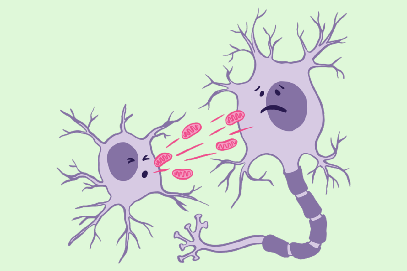
Individual technologies for recording and controlling neural activity in the brains of research mice have each advanced rapidly but the potential of easily mixing and matching them to conduct more sophisticated experiments, all while enabling the most natural behavior possible, has been difficult to realize. To empower a new generation of neuroscience experiments, engineers and scientists at MIT and the Open Ephys cooperative have developed a new standardized, open-source hardware and software platform. They described the system, called ONIX, in a new study Nov. 11 in Nature Methods.

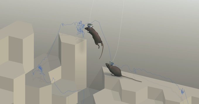
ONIX provides labs with a means to acquire data simultaneously from multiple popular implanted technologies (such as electrodes, microscopes and stimulation probes) while also powering and controlling those independent devices via a very thin coaxial cable and unimposing headstage. The system provides a standardized means of acquiring each instrument’s data and neatly integrating it all for efficient transmission to desktop software where scientists can then see and work with it. In the study the researchers document ONIX’s high data throughput and low latency. They also demonstrate that because the system’s headstage and cable are so physically light and resistant to twisting, mice can behave completely naturally and wear the system for days on end. In a large enclosure at MIT with a complex 3D landscape, for instance, mice wearing the system were able to nimbly scamper, climb and leap in experiments comparably to mice wearing no hardware at all.

“ONIX represents the culmination of many quantitative improvements that all come together to enable a qualitative leap in our ability to perform neural recordings in naturalistic behavior,” said corresponding author Jakob Voigts, an MIT neuroscience alumnus, co-founder of Open Ephys, and a research group leader at the Janelia Research Campus of the Howard Hughes Medical Institute. “We can now study the brain during behaviors that unfold over many hours and allow the animals to learn, to make a lot of complex decisions, and to interact with the world in ways that were previously not accessible.”

Jon Newman, a former MIT postdoc and now president of Open Ephys, and MIT postdoc Jie “Jack” Zhang led the work in the lab of co-author Matt Wilson, Sherman Fairchild Professor in The Picower Institute for Learning and Memory at MIT, together with Aarón Cuevas-López at Open Ephys. Wilson, whose lab studies neural processes underlying memory, said the idea behind developing ONIX was to develop a set of standards that would make it easy for any lab to use multiple technologies to acquire rich neural data while animals performed complex behaviors over long time periods.

“Jon’s motivation, the principle he used, was that if we need to do experiments that combined things like optogenetics, imaging, tetrode electrophysiology, and neuropixels, could we do it in a way that would not only enable experiments we were doing but also more complex experiments, involving more complex behavior, involving the integration of different recording methodologies that advances the whole community and not just one individual lab?,” said Wilson, a faculty member in MIT’s Departments of Biology and Brain and Cognitive Sciences (BCS).

Open origins

As Newman and then Zhang began to develop the technology starting in 2016 with this community-minded, open-source philosophy, Wilson said, it was natural to do so in partnership with Open Ephys, an MIT-born effort, now based in Atlanta, which develops and disseminates open, standardized systems to for neuroscience research. Making systems open-source provides researchers with many advantages, Voigts explained.

“Anyone can download the plans for the hardware as well as the software that make up the system,” Voigts said. “For technically well-versed neuroscientists this means that it is easier to modify aspects of the system. Open source also means that the system works with probes from many manufacturers because the connectors and standards aren’t proprietary. Most importantly, the open standards and design allow hardware and software developers to use ONIX as a starting point for completely new tools.”

Voigts compared ONIX to the USB standard people enjoy on their computers and phones. Any number of accessories can easily work with those devices because all they have to do is plug in. Similarly with ONIX, Wilson said, “You can mix and match and combine and then add new technologies without having to re-engineer the whole system.”

Lab demos

To validate the platform, the researchers conducted several experiments with mice including in Wilson’s lab and in the lab of co-author Mark Harnett, Associate Professor in the McGovern Institute for Brain Research and BCS Department at MIT (where Voigts did his postdoctoral work).

In their experiments they compared the mobility of mice implanted with electrodes but sometimes wearing ONIX (and its 0.3 mm tether cable) vs. sometimes wearing a commonly used and but substantially thicker (1.8 mm) tether cable over an 8-hour neural recording session. The mice proved to be much more mobile while wearing the lighter and thinner ONIX system, showing a broader range of exploration, freer head movement, and much faster running speeds. In a similar experiment in which mice were inplanted with tetrodes in the brain’s retrosplenial cortex, they even were able to jump while wearing ONIX but did not while wearing the more imposing tether. In another experiment the researchers compared mouse mobility around the enclosure between ONIX-wearing and completely unimplanted mice. The mice explored with equal freedom (as measured by motion tracking cameras) though the ONIX mice didn’t run as fast as unimplanted mice.

In further experiments, Voigts’s team at Janelia used ONIX to record for 55 hours because the system kept its cable tangle-free over that long-duration activity.

Finally the researchers showed that ONIX could transmit recordings not only from implanted electrodes and tetrodes but also from miniscopes and neuropixels, via experiments at the Allen Institute for Brain Science. They also showed how Open Ephys’s data acquisition software Bonsai (developed by co-author Goncalo Lopes) enabled the brain activity recordings to be synchronized with behavior tracking cameras to correlate neural activity and behavior.

Voigts said he hopes the system earns widespread adoption, especially as hardware costs continue to come down.

“I hope that this system convinces others to take the plunge and record neural data in more complex animal behaviors,” he said.

In addition to the authors named above, other authors are Nicholas Miller, Takato Honda, Marie-Sophie van der Goes, Alexandra Leighton Felipe Carvalho, Anna Lakunina, and Joshua Siegle, who co-founded Open Ephys with Voigts.

Funding for the study came from the National Institutes of Health, The Picower Institute for Learning and Memory, The JPB Foundation, the National Science Foundation, a Brain Science Foundation Research Grant Award, a Kavli-Grass-MBL Fellowship by the Kavli Foundation, the Grass Foundation, and Marine Biological Laboratory (MBL), an Osamu Hayaishi Memorial Scholarship for Study Abroad, a Uehara Memorial Foundation Overseas Fellowship, and Japan Society for the Promotion of Science (JSPS) Overseas Fellowship. a Mathworks Graduate Fellowship. The Simons Center for the Social Brain at MIT and the Howard Hughes Medical Institute.

A new approach to modeling complex biological systems

MIT engineers’ new model could help researchers glean insights from genomic data and other huge datasets. This is potentially critical to researchers who study any kind of complex biological system, according to senior author Douglas Lauffenburger.

Anne Trafton | MIT News
November 5, 2024

Over the past two decades, new technologies have helped scientists generate a vast amount of biological data. Large-scale experiments in genomics, transcriptomics, proteomics, and cytometry can produce enormous quantities of data from a given cellular or multicellular system.

However, making sense of this information is not always easy. This is especially true when trying to analyze complex systems such as the cascade of interactions that occur when the immune system encounters a foreign pathogen.

MIT biological engineers have now developed a new computational method for extracting useful information from these datasets. Using their new technique, they showed that they could unravel a series of interactions that determine how the immune system responds to tuberculosis vaccination and subsequent infection.

This strategy could be useful to vaccine developers and to researchers who study any kind of complex biological system, says Douglas Lauffenburger, the Ford Professor of Engineering in the departments of Biological Engineering, Biology, and Chemical Engineering.

“We’ve landed on a computational modeling framework that allows prediction of effects of perturbations in a highly complex system, including multiple scales and many different types of components,” says Lauffenburger, the senior author of the new study.

Shu Wang, a former MIT postdoc who is now an assistant professor at the University of Toronto, and Amy Myers, a research manager in the lab of University of Pittsburgh School of Medicine Professor JoAnne Flynn, are the lead authors of a new paper on the work, which appears today in the journal Cell Systems.

Modeling complex systems

When studying complex biological systems such as the immune system, scientists can extract many different types of data. Sequencing cell genomes tells them which gene variants a cell carries, while analyzing messenger RNA transcripts tells them which genes are being expressed in a given cell. Using proteomics, researchers can measure the proteins found in a cell or biological system, and cytometry allows them to quantify a myriad of cell types present.

Using computational approaches such as machine learning, scientists can use this data to train models to predict a specific output based on a given set of inputs — for example, whether a vaccine will generate a robust immune response. However, that type of modeling doesn’t reveal anything about the steps that happen in between the input and the output.

“That AI approach can be really useful for clinical medical purposes, but it’s not very useful for understanding biology, because usually you’re interested in everything that’s happening between the inputs and outputs,” Lauffenburger says. “What are the mechanisms that actually generate outputs from inputs?”

To create models that can identify the inner workings of complex biological systems, the researchers turned to a type of model known as a probabilistic graphical network. These models represent each measured variable as a node, generating maps of how each node is connected to the others.

Probabilistic graphical networks are often used for applications such as speech recognition and computer vision, but they have not been widely used in biology.

Lauffenburger’s lab has previously used this type of model to analyze intracellular signaling pathways, which required analyzing just one kind of data. To adapt this approach to analyze many datasets at once, the researchers applied a mathematical technique that can filter out any correlations between variables that are not directly affecting each other. This technique, known as graphical lasso, is an adaptation of the method often used in machine learning models to strip away results that are likely due to noise.

“With correlation-based network models generally, one of the problems that can arise is that everything seems to be influenced by everything else, so you have to figure out how to strip down to the most essential interactions,” Lauffenburger says. “Using probabilistic graphical network frameworks, one can really boil down to the things that are most likely to be direct and throw out the things that are most likely to be indirect.”

Mechanism of vaccination

To test their modeling approach, the researchers used data from studies of a tuberculosis vaccine. This vaccine, known as BCG, is an attenuated form of Mycobacterium bovis. It is used in many countries where TB is common but isn’t always effective, and its protection can weaken over time.

In hopes of developing more effective TB protection, researchers have been testing whether delivering the BCG vaccine intravenously or by inhalation might provoke a better immune response than injecting it. Those studies, performed in animals, found that the vaccine did work much better when given intravenously. In the MIT study, Lauffenburger and his colleagues attempted to discover the mechanism behind this success.

The data that the researchers examined in this study included measurements of about 200 variables, including levels of cytokines, antibodies, and different types of immune cells, from about 30 animals.

The measurements were taken before vaccination, after vaccination, and after TB infection. By analyzing the data using their new modeling approach, the MIT team was able to determine the steps needed to generate a strong immune response. They showed that the vaccine stimulates a subset of T cells, which produce a cytokine that activates a set of B cells that generate antibodies targeting the bacterium.

“Almost like a roadmap or a subway map, you could find what were really the most important paths. Even though a lot of other things in the immune system were changing one way or another, they were really off the critical path and didn’t matter so much,” Lauffenburger says.

The researchers then used the model to make predictions for how a specific disruption, such as suppressing a subset of immune cells, would affect the system. The model predicted that if B cells were nearly eliminated, there would be little impact on the vaccine response, and experiments showed that prediction was correct.

This modeling approach could be used by vaccine developers to predict the effect their vaccines may have, and to make tweaks that would improve them before testing them in humans. Lauffenburger’s lab is now using the model to study the mechanism of a malaria vaccine that has been given to children in Kenya, Ghana, and Malawi over the past few years.

“The advantage of this computational approach is that it filters out many biological targets that only indirectly influence the outcome and identifies those that directly regulate the response. Then it’s possible to predict how therapeutically altering those biological targets would change the response. This is significant because it provides the basis for future vaccine and trial designs that are more data driven,” says Kathryn Miller-Jensen, a professor of biomedical engineering at Yale University, who was not involved in the study.

Lauffenburger’s lab is also using this type of modeling to study the tumor microenvironment, which contains many types of immune cells and cancerous cells, in hopes of predicting how tumors might respond to different kinds of treatment.

The research was funded by the National Institute of Allergy and Infectious Diseases.

Sauer & Davis Lab News Brief: structures of molecular woodchippers reveal mechanism for versatility

Rest in pieces: deconstructing polypeptide degradation machinery

Lillian Eden | Department of Biology
November 12, 2024

Research from the Sauer and Davis Labs in the Department of Biology at MIT shows that conformational changes contribute to the specificity of “molecular woodchippers” 

Degradation is a crucial process for maintaining protein homeostasis by culling excess or damaged proteins whose components can then be recycled. It is also a highly regulated process—for good reason. A cell could potentially waste many resources if the degradation machinery destroys proteins it shouldn’t. 

One of the major pathways for protein degradation in bacteria and eukaryotic mitochondria involves a molecular machine called ClpXP. ClpXP is made up of two components: a star-shaped structure made up of six subunits called ClpX that engages and unfolds proteins tagged for degradation, and an associated barrel-shaped enzyme, called ClpP, that chemically breaks up proteins into small pieces called peptides. 

ClpXP is incredibly adaptable and is often compared to a woodchipper — able to take in materials and spit out their broken-down components. Thanks to biochemical experiments, this molecular degradation machine is known to be able to break down hundreds of different proteins in the cell regardless of physical or chemical properties such as size, shape, or charge. ClpX uses energy from ATP hydrolysis to unfold proteins before they are threaded through its central channel, referred to as the axial channel, and into the degradation chamber of ClpP.

In three papers, one in PNAS and two in Nature Communications, researchers from the Department of Biology at MIT have expanded our understanding of how this molecular machinery engages with, unfolds, and degrades proteins — and how that machinery refrains, by design, from unfolding proteins not tagged for degradation. 

Alireza Ghanbarpour, until recently a postdoc in the Sauer Lab and Davis Lab and first author on all three papers, began with a simple question: given the vast repertoire of potential substrates — that is, proteins to be degraded — how is ClpXP so specific?

Ghanbarpour — now an assistant professor in the Department of Biochemistry and Molecular Biology at Washington University School of Medicine in St. Louis — found that the answer to this question lies in conformational changes in the molecular machine as it engages with an ill-fated protein. 

Reverse Engineering using Structural Insights

Ghanbarpour approached the question of ClpXP’s versatility by characterizing conformational changes of the molecular machine using a technique called cryogenic electron microscopy. In cryo-EM, sample particles are frozen in solution, and images are collected; algorithms then create 3D renderings from the 2D images.

“It’s really useful to generate different structures in different conditions and then put them together until you know how a machine works,” he says. “I love structural biology, and these molecular machines make fascinating targets for structural work and biochemistry. Their structural plasticity and precise functions offer exciting opportunities to understand how nature leverages enzyme conformations to generate novel functions and tightly regulate protein degradation within the cell.”

Inside the cell, these proteases do not work alone but instead work together with “adaptor” proteins, which can promote — or inhibit — degradation by ClpXP. One of the adaptor proteins that promotes degradation by ClpXP is SspB. 

In E. coli and most other bacteria, ClpXP and SspB interact with a tag called ssrA that is added to incomplete proteins when their biosynthesis on ribosomes stalls. 

The tagging process frees up the ribosome to make more proteins, but creates a problem: incomplete proteins are prone to aggregation, which could be detrimental to cellular health and can lead to disease. By interacting with the degradation tag, ClpXP and SspB help to ensure the degradation of these incomplete proteins. Understanding this process and how it may go awry may open therapeutic avenues in the future.

“It wasn’t clear how certain adapters were interacting with the substrate and the molecular machines during substrate delivery,” Ghanbarpour notes. “My recent structure reveals that the adapter engages with the enzyme, reaching deep into the axial channel to deliver the substrate.” 

Ghanbarpour and colleagues showed that ClpX engages with both the SspB adaptor and the ssrA degradation tag of an ill-fated protein at the same time. Surprisingly, they also found that this interaction occurs while the upper part of the axial channel through ClpX is closed — in fact, the closed channel allows ClpX to contact both the tag and the adaptor simultaneously.

This result was surprising, according to senior author and Salvador E. Luria Professor of Biology Robert Sauer, whose lab has been working on understanding this molecular machine for more than two decades: it was unclear whether the channel through ClpX closes in response to a substrate interaction, or if the channel is always closed until it opens to pass an unfolded protein down to ClpP to be degraded.

Preventing Rogue Degradation

Throughout this project, Ghanbarpour was co-advised by structural biologist and Associate Professor of Biology Joey Davis and collaborated with members of the Davis Lab to better understand the conformational changes that allow these molecular machines to function. Using a cryo-EM analysis approach developed in the Davis lab called CryoDRGN, the researchers showed that there is an equilibrium between ClpXP in the open and closed states: it’s usually closed but is open in about 10% of the particles in their samples. 

The closed state is almost identical to the conformation ClpXP assumes when it is engaged with an ssrA-tagged substrate and the SspB adaptor. 

To better understand the biological significance of this equilibrium, Ghanbarpour created a mutant of ClpXP that is always in the open position. Compared to normal ClpXP, the mutant degraded some proteins lacking obvious degradation tags faster but degraded ssrA-tagged proteins more slowly. 

According to Ghanbarpour, these results indicate that the closed channel improves ClpXP’s ability to efficiently engage tagged proteins meant to be degraded, whereas the open channel allows more “promiscuous” degradation. 

Pausing the Process

The next question Ghanbarpour wanted to answer was what this molecular machine looks like while engaged with a protein it is attempting to unfold. To do that, he created a substrate with a highly stable protein attached to the degradation tag that is initially pulled into ClpX, but then dramatically slows protein unfolding and degradation.

In the structures where the degradation process stalls, Ghanbarpour found that the degradation tag was pulled far into the molecular machine—through ClpX and into ClpP—and the folded protein part of the substrate was pulled tightly against the axial channel of ClpX. 

The opening of the axial channel, called the axial pore, is made up of looping protein structures called RKH loops. These flexible loops were found to play roles both in recognizing the ssrA degradation tag and in how substrates or the SspB adaptor interact with or are pulled against the channel during degradation. 

The flexibility of these RKH loops allows ClpX to interact with a large number of different proteins and adapters, and these results clarify some previous biochemical and mutational studies of interactions between the substrate and ClpXP. 

Although Ghanbarpour’s recent work focused on just one adaptor and degradation tag, he noted there are many more targets — ClpXP is something akin to a Swiss army knife for breaking down polypeptide chains. 

The way those other substrates interact with ClpXP could differ from the structures solved with the SspB adaptor and ssrA tag. It also stands to reason that the way ClpXP reacts to each substrate may be unique. For example, given that ClpX is occasionally in an open state, some substrates may engage with ClpXP only while it’s in an open conformation. 

In his new position at Washington University, Ghanbarpour intends to continue exploring how ClpXP and other molecular machines locate their target substrates and interact with adaptors, shedding light on how cells regulate protein degradation and maintain protein homeostasis.

The structures Ghanbarpour solved involved free-floating protein degradation machinery, but membrane-bound degradation machinery also exists. The membrane-bound version’s structure and conformational adaptions potentially differ from the structures Ghanbarpour found in his previous three papers. Indeed, in a recent preprint, Ghanbarpour worked on the cryo-EM structure of a nautilus shell-shaped protein assembly that seems to control membrane-bound degradation machinery. This assembly plays a critical role in regulating protein degradation within the bacterial inner membrane.

“The function of these proteases goes beyond simply degrading damaged proteins. They also target transcription factors, regulatory proteins, and proteins that don’t exist in normal conditions,” he says. “My new lab is particularly interested in understanding how cells use these proteases and their accessory adaptors, both under normal and stress conditions, to reshape the proteome and support recovery from cellular distress.”

A cell protector collaborates with a killer

New research from the Horvitz Lab reveals what it takes for a protein that is best known for protecting cells against death to take on the opposite role.

Jennifer Michalowski | McGovern Institute
November 1, 2024

From early development to old age, cell death is a part of life. Without enough of a critical type of cell death known as apoptosis, animals wind up with too many cells, which can set the stage for cancer or autoimmune disease. But careful control is essential, because when apoptosis eliminates the wrong cells, the effects can be just as dire, helping to drive many kinds of neurodegenerative disease.

By studying the microscopic roundworm Caenorhabditis elegans—which was honored with its fourth Nobel Prize last month—scientists at MIT’s McGovern Institute have begun to unravel a longstanding mystery about the factors that control apoptosis: how a protein capable of preventing programmed cell death can also promote it. Their study, led by McGovern Investigator Robert Horvitz and reported October 9, 2024, in the journal Science Advances, sheds light on the process of cell death in both health and disease.

“These findings, by graduate student Nolan Tucker and former graduate student, now MIT faculty colleague, Peter Reddien, have revealed that a protein interaction long thought to block apoptosis in C. elegans, likely instead has the opposite effect,” says Horvitz, who shared the 2002 Nobel Prize for discovering and characterizing the genes controlling cell death in C. elegans.

Mechanisms of cell death

Horvitz, Tucker, Reddien and colleagues have provided foundational insights in the field of apoptosis by using C. elegans to analyze the mechanisms that drive apoptosis as well as the mechanisms that determine how cells ensure apoptosis happens when and where it should. Unlike humans and other mammals, which depend on dozens of proteins to control apoptosis, these worms use just a few. And when things go awry, it’s easy to tell: When there’s not enough apoptosis, researchers can see that there are too many cells inside the worms’ translucent bodies. And when there’s too much, the worms lack certain biological functions or, in more extreme cases, can’t reproduce or die during embryonic development.

Work in the Horvitz lab defined the roles of many of the genes and proteins that control apoptosis in worms. These regulators proved to have counterparts in human cells, and for that reason studies of worms have helped reveal how human cells govern cell death and pointed toward potential targets for treating disease.

A protein’s dual role

Three of C. elegans’ primary regulators of apoptosis actively promote cell death, whereas just one, CED-9, reins in the apoptosis-promoting proteins to keep cells alive. As early as the 1990s, however, Horvitz and colleagues recognized that CED-9 was not exclusively a protector of cells. Their experiments indicated that the protector protein also plays a role in promoting cell death. But while researchers thought they knew how CED-9 protected against apoptosis, its pro-apoptotic role was more puzzling.

CED-9’s dual role means that mutations in the gene that encode it can impact apoptosis in multiple ways. Most ced-9 mutations interfere with the protein’s ability to protect against cell death and result in excess cell death. Conversely, mutations that abnormally activate ced-9 cause too little cell death, just like mutations that inactivate any of the three killer genes.

An atypical ced-9 mutation, identified by Reddien when he was a PhD student in Horvitz’s lab, hinted at how CED-9 promotes cell death. That mutation altered the part of the CED-9 protein that interacts with the protein CED-4, which is proapoptotic. Since the mutation specifically leads to a reduction in apoptosis, this suggested that CED-9 might need to interact with CED-4 to promote cell death.

The idea was particularly intriguing because researchers had long thought that CED-9’s interaction with CED-4 had exactly the opposite effect: In the canonical model, CED-9 anchors CED-4 to cells’ mitochondria, sequestering the CED-4 killer protein and preventing it from associating with and activating another key killer, the CED-3 protein —thereby preventing apoptosis.

To test the hypothesis that CED-9’s interactions with the killer CED-4 protein enhance apoptosis, the team needed more evidence. So graduate student Nolan Tucker used CRISPR gene editing tools to create more worms with mutations in CED-9, each one targeting a different spot in the CED-4-binding region. Then he examined the worms. “What I saw with this particular class of mutations was extra cells and viability,” he says—clear signs that the altered CED-9 was still protecting against cell death, but could no longer promote it. “Those observations strongly supported the hypothesis that the ability to bind CED-4 is needed for the pro-apoptotic function of CED-9,” Tucker explains. Their observations also suggested that, contrary to earlier thinking, CED-9 doesn’t need to bind with CED-4 to protect against apoptosis.

When he looked inside the cells of the mutant worms, Tucker found additional evidence that these mutations prevented CED-9’s ability to interact with CED-4. When both CED-9 and CED-4 are intact, CED-4 appears associated with cells’ mitochondria. But in the presence of these mutations, CED-4 was instead at the edge of the cell nucleus. CED-9’s ability to bind CED-4 to mitochondria appeared to be necessary to promote apoptosis, not to protect against it.

Looking ahead

While the team’s findings begin to explain a long-unanswered question about one of the primary regulators of apoptosis, they raise new ones, as well. “I think that this main pathway of apoptosis has been seen by a lot of people as more or less settled science. Our findings should change that view,” Tucker says.

The researchers see important parallels between their findings from this study of worms and what’s known about cell death pathways in mammals. The mammalian counterpart to CED-9 is a protein called BCL-2, mutations in which can lead to cancer.  BCL-2, like CED-9, can both promote and protect against apoptosis. As with CED-9, the pro-apoptotic function of BCL-2 has been mysterious. In mammals, too, mitochondria play a key role in activating apoptosis. The Horvitz lab’s discovery opens opportunities to better understand how apoptosis is regulated not only in worms but also in humans, and how dysregulation of apoptosis in humans can lead to such disorders as cancer, autoimmune disease and neurodegeneration.

An elegant switch regulates production of protein variants during cell division

Cells make variants of thousands of proteins. These variants are not produced indiscriminately, but rather through precise regulatory mechanisms that can meet rapidly changing needs of the cell according to new research from the Cheeseman Lab.

Greta Friar | Whitehead Institute
October 18, 2024

Our cells contain thousands of proteins that have gone largely undetected and unstudied until recent years: these are variants of known proteins, which cells can make when their protein-building machinery interacts differently with the same stretch of genetic code. These protein variants have typically been overlooked as occasional accidents of gene expression, but researchers including Whitehead Institute Member Iain Cheeseman are discovering that they are actually abundant and can play important roles in cell functions. Researchers in Cheeseman’s lab are studying individual protein variants to learn more about them and their roles in health and disease, but they also wanted to understand broader patterns of protein variant production: how do cells control when to make one variant of a protein versus another, and what are the consequences of such switches?

Cheeseman, who is also a professor of biology at the Massachusetts Institute of Technology, and graduate student in his lab Jimmy Ly have now identified how cells switch to a different pattern of protein variant production during mitosis, or cell division. In research published in the journal Nature on October 23, they show that this broad regulatory switch helps cells survive paused cell divisions that can sometimes occur in healthy humans or be triggered by certain chemotherapy treatments. The work confirms that cells make variants of thousands of proteins, and also demonstrates that cells do not do so indiscriminately. Rather, cells use precise regulatory mechanisms to switch between different patterns of protein variant production, in order to rapidly tailor the proteins available to fit the changing needs of the cell.

A plethora of hidden proteins

Hw can our cells contain unknown proteins? In high school biology classes, students learn the rule that each gene codes for exactly one protein, such that if you know an organism’s genetic code, you should know every protein it can make. In fact, there are instead many genes that code for multiple proteins. For a protein to be made, first the genetic code for it is copied from DNA into a messenger RNA (mRNA). Then, a ribosome, the cellular machine that follows the instructions in genetic code to build a protein, locates the coding sequence within the mRNA by scanning for the start codon, a sequence of the three bases A, U, and G – bases are the chemical building blocks of RNA, abbreviated as A, U, C, and G. The ribosome recognizes the AUG start codon as the place to begin following instructions, and builds a protein based on the genetic sequence from there through to another trio of bases called a stop codon. However, one way that different versions of a protein can be produced is that a ribosome may begin reading the instructions from multiple different starting points.

Sometimes, a ribosome may miss the first AUG start codon and skip ahead to another AUG somewhere in the middle of the gene’s code, creating a truncated version of the protein. Sometimes, a ribosome may treat a similar trio of bases, such as CUG or GUG, as a start codon. This can cause it to begin earlier, creating a protein based on an extended genetic sequence. These possibilities mean that cells contain thousands more different proteins, or variants of proteins, than are represented by the dogma of one gene, one protein.

In order to understand protein variant production, the researchers—in collaboration with researchers from Whitehead Institute Member David Bartel’s lab–used a method that let them carefully track ribosomes to compare which start sites ribosomes tended to use. They looked at start site selection during mitosis versus during the rest of the cell cycle and found that a dramatic shift in use occurred for thousands of start sites. Specifically, the researchers found that during mitosis, ribosome scanning becomes more stringent. The ribosome will only begin making proteins at AUG sequences, and even then, only at AUGs that have preferable sequences of bases surrounding them—known as a strong Kozak context. This increased selectivity does not always lead to the familiar version of the protein being made during mitosis; sometimes the first AUG start codon has a weak Kozak context, so a truncated protein gets made from an AUG start codon with a stronger Kozak context that lies within the gene.

“Coming into this project, we knew very little about protein production during mitosis—for a long time, people didn’t think much protein production happened in mitosis at all,” Ly says. “It was satisfying to show not only that it is occurring, but that there’s a shift in which proteins are being made—and that this shift is important for cellular viability.”

How cells switch between protein variant programs

The researchers next identified how the switch to increased stringency is initiated during mitosis. They discovered that the key player is a protein called eIF1, which is one of many partners that can pair with ribosomes to help them select their start site. In particular, increased eIF1 pairing with ribosomes causes the ribosomes to be more stringent in their start codon selection, inhibiting the usage of non-AUG initiation sites or sites with weak Kozak contexts.

During mitosis, ribosome pairing with eIF1 increases sharply, leading to the shift in stringency. This change in pairing rate during mitosis puzzled the researchers: ribosomes and their partners, including eIF1, all typically reside together in the main body of the cell—where ribosomes make proteins—so they should be able to pair freely at any time. The researchers looked for other molecules in the same location that could be altering how ribosomes and eIF1 interact during different parts of the cell cycle, but they couldn’t find anything. Eventually, the researchers realized that the answer to the puzzle lay in a separate location: the nucleus.

They found that cells maintain a large pool of eIF1 inside of the nucleus, locked away from the ribosomes. Then, during cell division, the wall of the nucleus dissolves, mixing its contents with the rest of the cell. This is necessary for the dividing cell to divvy up its DNA, but it also releases the pool of eIF1 to pair with ribosomes, increasing stringency. At the end of mitosis, the nucleus reforms and eIF1 is re-incorporated into the nucleus of each of the two daughter cells, and the cells return to a less stringent program.

“The explanation for increased interaction between eIF1 and ribosomes during mitosis had really stumped us, and so when I saw eIF1 localizing to the nucleus, that was a really exciting ‘aha’ moment,” Ly says. “Discovering this mechanism of nuclear release during mitosis was unexpected, and it’s interesting to think about how else cells might be using it.”

Consequences of increased stringency for the cell

Once the researchers understood the how, they then wanted to understand the why? What they discovered is that when cells have no nuclear pool of eIF1, and so no change in stringency during mitosis, they are more likely to die during mitosis. In particular, these cells fare poorly during mitotic arrest, a state in which cells get stuck in mitosis for hours or even days–much longer than typical mitosis. Arrest occurs when cells detect a possible cell division error and so halt their division until the error is corrected or the cell dies.

One effect of increased stringency during mitosis is related to mitochondria, which are required for energy production in many cell types and are therefore required for maintaining viability. Cells stuck in mitotic arrest need energy to keep them going through this unexpected delay. The researchers found that increased stringency during mitosis led to an increase in the production of important mitochondrial proteins, boosting the cells’ energy supply to get them through arrest.

Increased stringency also gives cells the tools they need to escape arrest, even if they haven’t fixed the error that caused them to pause division. In a Nature paper in 2023, Cheeseman and then-postdoc in his lab Mary-Jane Tsang showed that when cells build up enough of the truncated version of a protein called CDC20, they can escape arrest. Ly’s work adds to this story by showing that the nuclear release of eIF1 increases stringency, leading to more production of truncated CDC20 during mitosis, which explains how cells build up enough of this protein variant during mitosis to trigger their escape. These findings may have important potential implications for some cancer chemotherapy strategies.

Some chemotherapies work by trapping cancer cells in mitotic arrest until they die. Cheeseman, Tsang, and Ly’s work collectively shows that when cancer cells lack sufficient truncated CDC20—as can occur in the absence of nuclear eIF1—the cells cannot escape arrest and so are killed off by these chemotherapies at higher rates. These results could be used to improve the efficacy of antimitotic chemotherapy drugs.

The switch in protein variant production that the researchers found affects thousands of proteins. These newly identified protein variants serve as a foundation for many future projects in the lab.

As the researchers continue to examine the consequences of this switch to stringency during mitosis, they are also searching for other cases in which cells regulate protein variant production outside of mitosis. For example, the researchers are interested in how this switch in stringency affects fertility; immature egg cells spend a long time in a form of arrested cell division without an intact nucleus, and Ly observed eIF1 in the nucleus of the immature female eggs.

“Cells have axes of control that they use to quickly make broad changes in gene expression,” Cheeseman says. “Several of these are central to controlling cell division—for example, the role of phosphorylation as a regulatory switch in mitosis has been well studied. Our work identifies another axis of control, and we’re excited to discover more about when and how cells make use of it.”

Laub Lab News Brief: anti-viral defense system in bacteria modifies mRNA

Killing the messenger

Lillian Eden | Department of Biology
October 23, 2024

Newly characterized anti-viral defense system in bacteria aborts infection through novel mechanism by chemically modifying mRNA.


Like humans and other complex multicellular organisms, single-celled bacteria can fall ill and fight off viral infections. A bacterial virus is known as a bacteriophage, or, more simply, a phage, which is one of the most ubiquitous life forms on Earth. Phages and bacteria are engaged in a constant battle, the virus attempting to circumvent the bacteria’s defenses, and the bacteria racing to find new ways to protect itself.

These anti-phage defense systems are carefully controlled and prudently managed — dormant but always poised to strike. 

New research recently published in Nature from the Laub Lab in the Department of Biology at MIT has characterized an anti-phage defense system in bacteria known as CmdTAC. CmdTAC prevents viral infection by altering mRNA, the single-stranded genetic code used to produce proteins, of both the host and the virus.  

This defense system detects phage infection at a stage when the viral phage has already commandeered the host’s machinery for its own purposes. In the face of annihilation, the ill-fated bacterium activates a defense system that will halt translation, preventing the creation of new proteins and aborting the infection — but dooming itself in the process. 

“When bacteria are in a group, they’re kind of like a multicellular organism that is not connected to one another. It’s an evolutionarily beneficial strategy for one cell to kill itself to save another identical cell,” says Christopher Vassallo, a postdoc and co-author of the study. “You could say it’s like self-sacrifice: one cell dies to protect the other cells.” 

The enzyme responsible for altering the mRNA is called an ADP-ribosyltransferase.  Researchers have characterized hundreds of these enzymes — although only a few are known to target DNA or other types of RNA, all but a handful target proteins. This is the first time these enzymes have been characterized targeting mRNA within cells.

Expanding understanding of anti-phage defense

Co-first author and graduate student Chris Doering noted that it is only within the last decade or so that researchers have begun to appreciate the breadth of diversity and complexity of anti-phage defense systems. For example, CRISPR gene editing, a technique used in everything from medicine to agriculture, is rooted in research on the bacterial CRISPR-Cas9 anti-phage defense system. 

CmdTAC is a subset of a widespread anti-phage defense mechanism called a toxin-antitoxin system. A TA system is just that: a toxin capable of killing or altering the cell’s processes rendered inert by an associated antitoxin. 

Although these TA systems can be identified — if the toxin is expressed by itself, it kills or inhibits the growth of the cell; if the toxin and antitoxin are expressed together, the toxin is neutralized — characterizing the cascade of circumstances that activates these systems requires extensive effort. In recent years, however, many TA systems have been shown to serve as anti-phage defenses. 

Two general questions need to be answered to understand a viral defense system: how do bacteria detect an infection, and how do they respond?

Detecting infection

CmdTAC is a TA system with an additional element, and the three components generally exist in a stable complex: the toxin CmdT, the antitoxin CmdA, and an additional component that mediates the system, the chaperone CmdC. 

If the phage’s protective capsid protein is present, CmdC disassociates from CmdT and CmdA and interacts with the phage capsid protein instead. In the model outlined in the paper, the chaperone CmdC is, therefore, the sensor of the system, responsible for recognizing when an infection is occurring. Structural proteins, such as the capsid that protects the phage genome, are a common trigger because they’re abundant and essential to the phage.

The uncoupling of CmdC leads to the degradation of the neutralizing antitoxin CmdA, which releases the toxin CmdT to do its lethal work.

Toxicity on the loose

Guided by computational tools, the researchers knew that CmdT was likely an ADP-ribosyltransferase due to its similarities to other such enzymes. As the name suggests, the enzyme transfers an ADP ribose onto its target.

To determine how CmdT was altering mRNA, the researchers tested a mix of short sequences of single-stranded RNA to see if the enzyme was drawn to any sequences or positions in particular. RNA has four bases: A, U, G, and C, and the evidence points to the enzyme recognizing GA sequences. 

The CmdT modification of GA sequences in mRNA blocks its translation. The cessation of creating new proteins aborts the infection, preventing the phage from spreading beyond the host to infect other bacteria. 

“Not only is it a new type of bacterial immune system, but the enzyme involved does something that’s never been seen before: the ADP-ribsolyation of mRNA,” Vassallo says. 

Although the paper outlines the broad strokes of the anti-phage defense system, there’s more to learn: it’s unclear how CmdC interacts with the capsid protein, and how the chemical modification of GA sequences prevents translation. 

Beyond Bacteria

While exploring anti-phage defense aligns with the Laub Lab’s overall goal of understanding how bacteria function and evolve, these results may have broader implications beyond bacteria.

Senior author Michael Laub, Salvador E. Luria Professor and HHMI Investigator, says the ADP-ribosyltransferase has homologs in eukaryotes, including human cells. They are not well studied, and not currently among the Laub Lab’s research topics, but they are known to be up-regulated in response to viral infection. 

“There are so many different — and cool — mechanisms by which organisms defend themselves against viral infection,” Laub says. “The notion that there may be some commonality between how bacteria defend themselves and how humans defend themselves is a tantalizing possibility.” 

Establishing boundaries of the genetic kind

The pseudoautosomal region (PAR) is a critical area on the Y chromosome that swaps genetic information with the X chromosome. Recent research from the Page Lab reaffirms the location of PAR and offers a refined understanding of where crossover events occur.

Shafaq Zia | Whitehead Institute
October 14, 2024

At first, the X and the Y sex chromosomes seemed like an unlikely pair. But then, researchers, including Whitehead Institute Member David Page, began finding clues that suggested otherwise: identical DNA sequences on the X and Y chromosomes.

Soon, it became clear that the tips of the X and Y chromosomes join together in a tight embrace, swapping genetic material during the process of sperm production from immature male germ cells. This limited area of genetic exchange between the two sex chromosomes is called the pseudoautosomal region (PAR).

But science is an iterative process—a continuous cycle of questioning, testing, and revising knowledge. Last fall, what had long been considered well established in genetics was called into question when new research suggested that the boundary of the PAR might be half a million base pairs away from the accepted location. Given that a typical human gene is about tens of thousands of base pairs, this length would potentially span multiple genes on the X and Y chromosomes, raising serious concerns about the accuracy and validity of decades of scientific literature.

Fortunately, new work from Page, research scientist Daniel Winston Bellott, and colleagues—published Oct. 14 in the American Journal of Human Genetics—offers clarity. In this study, the group re-examines the size of the PAR using sequencing data presented by outside researchers in their 2023 work, alongside decades of genomic resources, and single-cell sequencing of human sperm. Their findings confirm that the location of the boundary to the PAR, as identified by scientists in 1989, still holds true.

“If one is interested in understanding sex differences in health and disease, the boundary of the pseudoautosomal region is arguably the most fundamental landmark in the genome,” says Page, who is also a professor of biology at the Massachusetts Institute of Technology and an Investigator with Howard Hughes Medical Institute. “Had this boundary been multiple genes off, the field would have been shaken to its foundations.”

Dance of the chromosomes

The X and Y chromosomes evolved from an ancestral pair of chromosomes with identical structures. Over time, the Y chromosome degenerated drastically, losing hundreds of functional genes. Despite their differences, the X and Y chromosomes come together during a special type of cell division called male meiosis, which produces sperm cells.

This process begins with the tips of the sex chromosomes aligning side by side like two strands of rope. As the X and Y chromosomes embrace each other, enzymes create breaks in the DNA. These breaks are repaired using the opposite chromosome as a template, linking the X and Y together. About half of the time, an entire segment of DNA, which often contains multiple genes, will cross over onto the opposite chromosome.

The genetic exchange, called recombination, concludes with the X and Y chromosomes being pulled apart to opposite ends of the dividing cell, ensuring that each chromosome ends up in a different daughter cell. “This intricate dance of the X and Y chromosomes is essential to a sperm getting either an X or a Y—not both, and not neither,” says Page.

This way when the sperm—carrying either an X or a Y—fuses with the egg—carrying an X—during fertilization, the resulting zygote has the right number of chromosomes and a mix of genetic material from both parents.

But that’s not all. The swapping of DNA during recombination also allows for the chromosomes to have the same genes but with slight variations. These unique combinations of genetic material across sex chromosomes are key to genetic diversity within a species, enabling it to survive, adapt, and reproduce successfully.

Beyond the region of recombination, the Y chromosome contains genes that are important for sex determination, for sperm production, and for general cellular functioning. The primary sex-defining gene, SRY, which triggers the development of an embryo into a male, is located only 10,000 bases from the boundary of the PAR.

Advancing together

To determine whether the location of this critical boundary on the human sex chromosomes—where they stop crossing over during meiosis and become X-specific or Y-specific—had been misidentified for over three decades, researchers began by comparing publicly-available DNA sequences from the X and the Y chromosomes of seven primate species: humans, chimpanzees, gorillas, orangutans, siamangs, rhesus macaques, and colobus monkeys.

Based on the patterns of crossover between the X and the Y chromosomes of these species, the researchers constructed an evolutionary tree. Upon analyzing how DNA sequences close to and distant from the PAR boundary group together across species, the researchers found a substitution mutation—where a letter in a long string of letters is swapped for a different one—in the DNA of the human X and Y chromosomes. This change was also present in the chimpanzee Y chromosome, suggesting that the mutation originally occurred in the last common ancestor of humans and chimpanzees and was then transferred to the human X chromosome.

“These alignments between various primates allowed us to observe where the X and the Y chromosomes have preserved identity over millions of years and where they have diverged,” says Bellott. “That [pseudoautosomal] boundary has remained unchanged for 25 million years.”

Next, the group studied crossover events in living humans using a vast dataset of single-cell sequencing of sperm samples. They found 795 sperm with clear swapping of genetic material somewhere between the originally proposed boundary of the PAR and the newly-proposed 2023 boundary.

Once these analyses confirmed that the original location of the PAR boundary remains valid, Page and his team turned their attention to data from the 2023 study that contested this 1989 finding. The researchers focused on 10 male genomes assembled by the outside group, which contained contiguous sequences from the PAR.

Since substitutions on the Y chromosome typically occur at a steady rate, but in the PAR, changes on the X chromosome can transfer to the Y through recombination, the researchers compared the DNA sequences from the ten genomes to determine whether they followed the expected steady rate of change or if they varied.

The team found that close to the originally proposed PAR boundary, the DNA sequences changed at a steady rate. But further away from the boundary, the rate of change varied, suggesting that crossover events likely occurred in this region. Furthermore, the group identified several shared genetic differences between the X and the Y chromosomes of these genomes, which demonstrates that recombination has occurred even closer to the PAR boundary than scientists observed in 1989.

“Ironically, instead of contradicting the original boundary, the 2023 work has helped us refine the location of crossover to an even narrower area near the boundary,” says Page.

Thanks to the efforts of Page’s group at Whitehead Institute, our understanding of the PAR is clearer than ever, and business can go on as usual for researchers investigating sex differences in health and disease.

Bat cells possess a unique antiviral mechanism, preventing the SARS-CoV-2 virus from taking control

Bats have the amazing ability to coexist with viruses that are deadly to humans. New work from the Jaenisch Lab uncovers an antiviral mechanism that allows viruses to enter bat cells but prevents them from replicating.

Shafaq Zia | Whitehead Institute
October 14, 2024

Viruses are masters of stealth. From the moment a virus enters the host’s body, it begins hijacking its cells. First, the virus binds to a specific protein on the cell’s surface through a lock-and-key mechanism. This protein, known as a receptor, facilitates the entry of the virus’s genetic material into the cell. Once inside, this genetic code takes over the cell’s machinery, directing it to produce copies of the virus and assemble new viral particles, which can go on to infect other cells. Upon detecting the invasion, the host’s immune system responds by attacking infected cells in hopes of curbing the virus’s spread.

But in bats, this process unfolds differently. Despite carrying several viruses — Marburg, Ebola, Nipah, among others — bats rarely get sick from these infections. It seems their immune systems are highly specialized, allowing them to live with viruses that would typically be deadly in humans, without any clinical symptoms.

Since the onset of the COVID-19 pandemic, the lab of Whitehead Institute Founding Member Rudolf Jaenisch has been investigating the molecular basis of bats’ extraordinary resilience to viruses like SARS-CoV-2. In their latest study, published in the journal PNAS on Oct. 14 , Jaenisch lab postdoc Punam Bisht and colleagues have uncovered an antiviral mechanism in bat cells that allows viruses to enter the cells but prevents them from replicating their genome and completing the hijacking process.

“These cells have elevated expressions of antiviral genes that act immediately, neutralizing the virus before it can spread,” says Jaenisch, a professor of biology at the Massachusetts Institute of Technology. “What’s particularly interesting is that many of these antiviral genes have counterparts, or orthologs in humans.”

Striking a delicate balance

The innate immune system is the body’s first line of defense against foreign invaders like the SARS-CoV-2 virus. This built-in security system is always on alert, responding swiftly — within minutes to hours — to perceived threats.

Upon detecting danger, immune cells rush to the site of infection, where they target the virus with little precision in attempts to slow it down and buy time for the more specialized adaptive immune system to take over. During this process, these cells release small signaling proteins called cytokines, which coordinate the immune response by recruiting additional immune cells and directing them to the battleground.

If the innate immune response alone isn’t sufficient to defeat the virus, it signals the adaptive immune system for support. The adaptive immune system tailors its attacks to the exact pathogen it is fighting and can even keep records of past infections to launch a faster, more aggressive attack the next time it encounters the same pathogen.

But in some infections, the innate immune response can quickly spiral out of control before the adaptive immune response is activated. This phenomenon, called a cytokine storm, is a life-threatening condition characterized by the overproduction of cytokines. These proteins continue to signal the innate immune system for backup even when it’s not necessary, leading to a flood of immune cells at the site of infection, where they inadvertently begin damaging organs and healthy tissues.

Bats, on the other hand, are uniquely equipped to manage viral infections without triggering an overwhelming immune response or allowing the virus to take control. To understand how their innate immune system achieves this delicate balance, Bisht and her colleagues turned their attention to bat cells.

In this study, researchers compared how the SARS-CoV-2 virus replicates in human and bat stem cells and fibroblasts — a type of cell involved in the formation of connective tissue. While fibroblasts are not immune cells, they can secrete cytokines and guide immune response, particularly to help with tissue repair.

After exposing these cells to the SARS-CoV-2 virus for 48 hours, the researchers used a Green Fluorescent Protein (GFP) tag to track the virus’s activity. GFP is a fluorescent protein whose genetic code can be added as a tag to a gene of interest. This causes the products of that gene to glow, providing researchers with a visual marker of where and when the gene is expressed.

They observed that over 80% of control cells — derived from the kidneys of African green monkeys and known to be highly susceptible to SARS-CoV-2 — showed evidence of the virus replicating. In contrast, they did not detect any viral activity in human and bat stem cells or fibroblasts.

In fact, even after introducing the human ACE2 receptor — which SARS-CoV-2 uses to bind and enter cells — into bat cells, the infected bat fibroblasts were able to replicate viral RNA and produce viral proteins, but at much lower levels compared to infected human fibroblasts.

These bat fibroblasts, however, could not assemble these viral proteins into fully infectious virus particles, suggesting an abortive infection, where the virus is able to initiate replication but fails to complete the process and produce progeny viruses.

Using electron microscopy to look inside bat and human cells, they began to understand why: in human cells, SARS-CoV-2 had created special structures called double-membrane vesicles (DMV). These vesicles acted like a bubble, shielding the viral genome from detection and providing it safe space to replicate more effectively. However, these “viral replication factories” were absent in bat fibroblasts.

When the researchers examined the gene expression profiles of these bat fibroblasts and compared them those of infected human cells, they found that although both human and bat cells have genes regulating the release of a type of cytokine called interferons, these genes are already turned on in bat fibroblasts — unlike in human cells — even before virus infection occurs.

These findings suggest that bat cells are in a constant state of vigilance. This allows their innate immune system to stop the SARS-CoV-2 virus in its tracks early on in the replication process before it can entirely hijack cellular machinery.

Surprisingly, this antiviral mechanism does not protect bat cells against all viruses. When the researchers infected bat fibroblasts with Zika virus, the virus was able to replicate and produce new viral particles.

“This means there are still many questions unanswered about how bat cells resist infection,” says Bisht. “COVID-19 continues to circulate, and the virus is evolving quickly. Filling in these gaps in our knowledge will help us develop better vaccines and antiviral strategies.”
The researchers are now focused on identifying the specific genes involved in this antiviral mechanism, and exploring how they interact with the virus during infection.

Cancer biologists discover a new mechanism for an old drug

Study reveals the drug, 5-fluorouracil, acts differently in different types of cancer — a finding that could help researchers design better drug combinations.

Anne Trafton | MIT News
October 7, 2024

Since the 1950s, a chemotherapy drug known as 5-fluorouracil has been used to treat many types of cancer, including blood cancers and cancers of the digestive tract.

Doctors have long believed that this drug works by damaging the building blocks of DNA. However, a new study from MIT has found that in cancers of the colon and other gastrointestinal cancers, it actually kills cells by interfering with RNA synthesis.

The findings could have a significant effect on how doctors treat many cancer patients. Usually, 5-fluorouracil is given in combination with chemotherapy drugs that damage DNA, but the new study found that for colon cancer, this combination does not achieve the synergistic effects that were hoped for. Instead, combining 5-FU with drugs that affect RNA synthesis could make it more effective in patients with GI cancers, the researchers say.

“Our work is the most definitive study to date showing that RNA incorporation of the drug, leading to an RNA damage response, is responsible for how the drug works in GI cancers,” says Michael Yaffe, a David H. Koch Professor of Science at MIT, the director of the MIT Center for Precision Cancer Medicine, and a member of MIT’s Koch Institute for Integrative Cancer Research. “Textbooks implicate the DNA effects of the drug as the mechanism in all cancer types, but our data shows that RNA damage is what’s really important for the types of tumors, like GI cancers, where the drug is used clinically.”

Yaffe, the senior author of the new study, hopes to plan clinical trials of 5-fluorouracil with drugs that would enhance its RNA-damaging effects and kill cancer cells more effectively.

Jung-Kuei Chen, a Koch Institute research scientist, and Karl Merrick, a former MIT postdoc, are the lead authors of the paper, which appears today in Cell Reports Medicine.

An unexpected mechanism

Clinicians use 5-fluorouracil (5-FU) as a first-line drug for colon, rectal, and pancreatic cancers. It’s usually given in combination with oxaliplatin or irinotecan, which damage DNA in cancer cells. The combination was thought to be effective because 5-FU can disrupt the synthesis of DNA nucleotides. Without those building blocks, cells with damaged DNA wouldn’t be able to efficiently repair the damage and would undergo cell death.

Yaffe’s lab, which studies cell signaling pathways, wanted to further explore the underlying mechanisms of how these drug combinations preferentially kill cancer cells.

The researchers began by testing 5-FU in combination with oxaliplatin or irinotecan in colon cancer cells grown in the lab. To their surprise, they found that not only were the drugs not synergistic, in many cases they were less effective at killing cancer cells than what one would expect by simply adding together the effects of 5-FU or the DNA-damaging drug given alone.

“One would have expected that these combinations to cause synergistic cancer cell death because you are targeting two different aspects of a shared process: breaking DNA, and making nucleotides,” Yaffe says. “Karl looked at a dozen colon cancer cell lines, and not only were the drugs not synergistic, in most cases they were antagonistic. One drug seemed to be undoing what the other drug was doing.”

Yaffe’s lab then teamed up with Adam Palmer, an assistant professor of pharmacology at the University of North Carolina School of Medicine, who specializes in analyzing data from clinical trials. Palmer’s research group examined data from colon cancer patients who had been on one or more of these drugs and showed that the drugs did not show synergistic effects on survival in most patients.

“This confirmed that when you give these combinations to people, it’s not generally true that the drugs are actually working together in a beneficial way within an individual patient,” Yaffe says. “Instead, it appears that one drug in the combination works well for some patients while another drug in the combination works well in other patients. We just cannot yet predict which drug by itself is best for which patient, so everyone gets the combination.”

These results led the researchers to wonder just how 5-FU was working, if not by disrupting DNA repair. Studies in yeast and mammalian cells had shown that the drug also gets incorporated into RNA nucleotides, but there has been dispute over how much this RNA damage contributes to the drug’s toxic effects on cancer cells.

Inside cells, 5-FU is broken down into two different metabolites. One of these gets incorporated into DNA nucleotides, and other into RNA nucleotides. In studies of colon cancer cells, the researchers found that the metabolite that interferes with RNA was much more effective at killing colon cancer cells than the one that disrupts DNA.

That RNA damage appears to primarily affect ribosomal RNA, a molecule that forms part of the ribosome — a cell organelle responsible for assembling new proteins. If cells can’t form new ribosomes, they can’t produce enough proteins to function. Additionally, the lack of undamaged ribosomal RNA causes cells to destroy a large set of proteins that normally bind up the RNA to make new functional ribosomes.

The researchers are now exploring how this ribosomal RNA damage leads cells to under programmed cell death, or apoptosis. They hypothesize that sensing of the damaged RNAs within cell structures called lysosomes somehow triggers an apoptotic signal.

“My lab is very interested in trying to understand the signaling events during disruption of ribosome biogenesis, particularly in GI cancers and even some ovarian cancers, that cause the cells to die. Somehow, they must be monitoring the quality control of new ribosome synthesis, which somehow is connected to the death pathway machinery,” Yaffe says.

New combinations

The findings suggest that drugs that stimulate ribosome production could work together with 5-FU to make a highly synergistic combination. In their study, the researchers showed that a molecule that inhibits KDM2A, a suppressor of ribosome production, helped to boost the rate of cell death in colon cancer cells treated with 5-FU.

The findings also suggest a possible explanation for why combining 5-FU with a DNA-damaging drug often makes both drugs less effective. Some DNA damaging drugs send a signal to the cell to stop making new ribosomes, which would negate 5-FU’s effect on RNA. A better approach may be to give each drug a few days apart, which would give patients the potential benefits of each drug, without having them cancel each other out.

“Importantly, our data doesn’t say that these combination therapies are wrong. We know they’re effective clinically. It just says that if you adjust how you give these drugs, you could potentially make those therapies even better, with relatively minor changes in the timing of when the drugs are given,” Yaffe says.

He is now hoping to work with collaborators at other institutions to run a phase 2 or 3 clinical trial in which patients receive the drugs on an altered schedule.

“A trial is clearly needed to look for efficacy, but it should be straightforward to initiate because these are already clinically accepted drugs that form the standard of care for GI cancers. All we’re doing is changing the timing with which we give them,” he says.

The researchers also hope that their work could lead to the identification of biomarkers that predict which patients’ tumors will be more susceptible to drug combinations that include 5-FU. One such biomarker could be RNA polymerase I, which is active when cells are producing a lot of ribosomal RNA.

The research was funded by the Damon Runyon Cancer Research Fund, a Ludwig Center at MIT Fellowship, the National Institutes of Health, the Ovarian Cancer Research Fund, the Holloway Foundation, and the STARR Cancer Consortium.

Brain cell types are affected differently by Rett Syndrome mutation

New research from Jaenisch Lab postdoc Danielle Tomasello focuses on an understudied question: how Rett Syndrome affects cell types in the human brain other than neurons.

Greta Friar | Whitehead Institute
September 6, 2024

Rett Syndrome is a X-chromosome-linked neurodevelopmental disorder; it can lead to loss of coordination, mobility, ability to speak, and use of the hands, among other symptoms. The syndrome is typically caused by mutations within the gene MECP2. Researchers in Whitehead Institute Founding Member Rudolf Jaenisch’s lab have studied Rett Syndrome for many years in order to understand the biological mechanisms that cause disease symptoms, and to identify possible avenues for treatments or a cure. Jaenisch and colleagues have gained many insights into the biology of Rett syndrome and developed tools that can rescue neurons from Rett syndrome symptoms in lab models.

However, much about the biology of Rett Syndrome remains unknown. New research from Jaenisch and postdoc in his lab Danielle Tomasello focuses on an understudied question: how Rett Syndrome affects cell types in the human brain other than neurons. Specifically, Tomasello investigated the effects of Rett Syndrome on astrocytes, a type of brain cell that supports and provides energy for neurons. The work, shared in the journal Scientific Reports on September 6, details changes that occur in Rett syndrome astrocytes, in particular in relation to their mitochondria, and shows how these changes directly impact neurons. The findings provide a new framework for thinking about Rett Syndrome and possible new avenues for therapies.

“By considering Rett Syndrome from a different perspective, this project expands our understanding of a multifaceted and thus far incurable disease,” says Jaenisch, who is also a professor of biology at the Massachusetts Institute of Technology.

Energy metabolism in Rett Syndrome

Mitochondria are organelles that generate energy, which cells use to carry out their functions, and mitochondrial dysfunction was known to occur in Rett Syndrome. Jaenisch and Tomasello found that mitochondria in astrocytes are particularly affected, even more so than mitochondria in neurons. Tomasello grew human stem-cell-derived astrocytes in 2D cultures and also grew 3D organoids: mini brain-like tissues that contain multiple cell types growing in a structure that resembles actual brain anatomy. This approach allowed Tomasello to use human cells, rather than an animal model, and to study how cells behave within a brain-like environment.

When the researchers observed Rett astrocytes grown in these conditions, they found that the mitochondria were misshapen: short, small circles instead of large, long ovals. Additional studies showed evidence of the mitochondria experiencing stress and not being able to generate enough energy through their usual processes. The mitochondria did not have enough of the typical proteins they use to make energy, and so began to break down the cell’s supply of the building blocks of proteins, amino acids, for parts to make up for the missing material. Additionally, the researchers observed an increase in reactive oxygen species, byproducts of mitochondrial metabolism that are toxic to the cell.

Further experiments suggested that the cells try to compensate for this mitochondrial stress by increasing transcription of mitochondrial genes. For example, Tomasello found that regions of DNA called promoters that can increase expression of key mitochondrial genes were more open for the cell to use in Rett astrocytes. Altogether, these findings paint a picture of severe mitochondrial dysfunction in Rett astrocytes.

Although mitochondria in Rett neurons did not have such severe defects, astrocytes and neurons have a close relationship. Not only do neurons rely on astrocytes to supply them with energy, they even accept mitochondria from astrocytes to use for themselves. Jaenisch and Tomasello found that neurons take up dysfunctional mitochondria from Rett astrocytes at a higher rate than they take up mitochondria from unaffected astrocytes. This means that the effects of Rett syndrome on astrocytes have a direct effect on neurons: the dysfunctional mitochondria from the astrocytes end up in the neurons, where they cause damage. Tomasello took mitochondria from Rett astrocytes and placed them on both healthy and Rett neurons. In either case, the neurons took up the dysfunctional mitochondria in large numbers and then experienced significant problems. The neurons entered a hyperexcitable state that is ultimately toxic to the brain. The neurons also contained higher levels of reactive oxygen species, the toxic byproducts of mitochondrial metabolism, which can cause widespread damage. These effects occurred even in otherwise healthy neurons that did not themselves contain a Rett-causing MECP2 mutation.

“This shows that in order to understand Rett Syndrome, we need to look beyond what’s happening in neurons to other cell types,” Tomasello says.

Learning about the role that astrocytes play in Rett Syndrome could provide new avenues for therapies. The researchers found that supplying affected astrocytes with healthy mitochondria helped them to recover normal mitochondrial function. This suggests to Tomasello that one possibility for future Rett Syndrome therapies could be something that either targets mitochondria, or supplies additional mitochondria through the bloodstream.

Together, these insights and their possible medical implications demonstrate the importance of taking a broader look at the foundational biology underlying a disease.